Durable survival with OPDIVO ® (nivolumab) + chemotherapy (fluoropyrimidine- and platinum-based) vs chemotherapy alone, a first-line treatment of metastatic gastric cancer, gastroesophageal junction cancer, and esophageal adenocarcinoma, regardless of PD-L1 status at 4 years of follow-up1,2

Ronan Kelly, MD, MBA,
The Charles A. Sammons Cancer Center,
Baylor University Medical Center, Dallas, Texas*
Content sponsored by: Bristol Myers Squibb
*Dr Kelly was compensated by BMS for his contribution in drafting this article.

Introduction: Overview of gastroesophageal adenocarcinoma
Gastroesophageal adenocarcinomas consist of a heterogeneous group of tumors, including gastric cancer (GC), gastroesophageal junction cancer (GEJC), and esophageal adenocarcinoma (EAC), all of which are aggressive malignancies with poor outcomes.3-6 The aggressive natures of GC and EAC may contribute to their respective statuses as two of the most common causes of cancer-related death globally.7
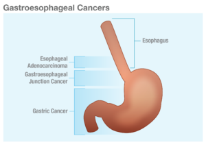
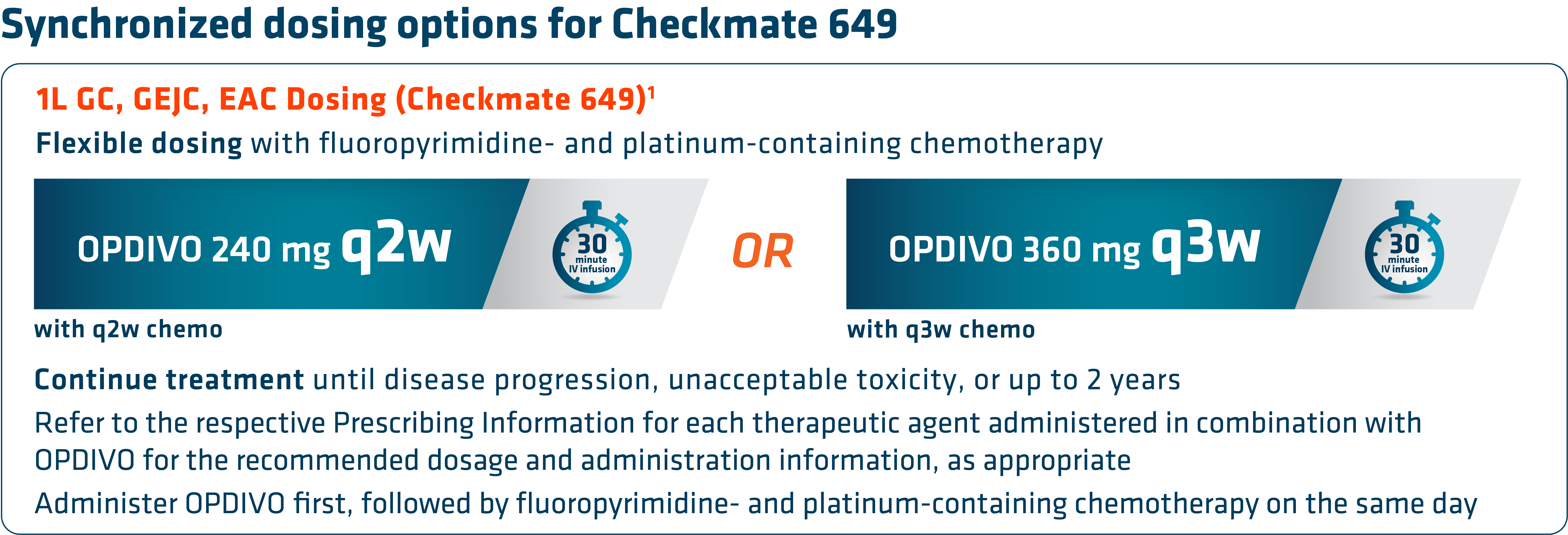
Gastroesophageal-CancersCheckmate 649 led to the approval of nivolumab (OPDIVO) + chemotherapy as the first chemoimmunotherapy combination for all eligible patients with HER2-negative GC/GEJC/EAC regardless of PD-L1 status.1,8,9 Prior to this approval, chemotherapy was the only available 1L treatment option for metastatic GC/GEJC/EAC.10 Furthermore, to date, Checkmate 649 has the longest follow-up survival data in GC vs chemotherapy for any I-O–based regimen with a minimum follow-up of 48.1 months (median of 59.3 months), and showed durable survival data with OPDIVO + chemotherapy in GC/GEJC/EAC.1,2,11 OPDIVO can be given q2w or q3w, which synchronizes with the q2w FOLFOX and q3w CapeOx dosing schedules.1 “The flexible dosing schedule of OPDIVO has made it more convenient to integrate into my clinical practice,” stated Dr. Kelly.

Indication has no restriction on HER2 status; trial included HER2-negative patients and patients with unknown HER2 status, while excluding those with known HER2-positive status.1

OPDIVO + chemotherapy in 1L metastatic GC/GEJC/EAC
With the longest follow-up survival data in GC vs chemotherapy for any I-O–based regimen and durable survival data in GC/GEJC/EAC, OPDIVO + fluoropyrimidine- and platinum-containing chemotherapy is currently FDA-approved in 1L metastatic non–HER2-positive GC/GEJC/EAC, regardless of PD-L1 status (no testing required).1,2,9 The approval of this combination was based on the results of Checkmate 649, a global phase 3 study in patients with 1L metastatic GC/GEJC/EAC.1,8 Key exclusion criteria included known HER2-positive status and untreated CNS metastases.8 The study recruited all eligible patients regardless of PD-L1 expression.1,8
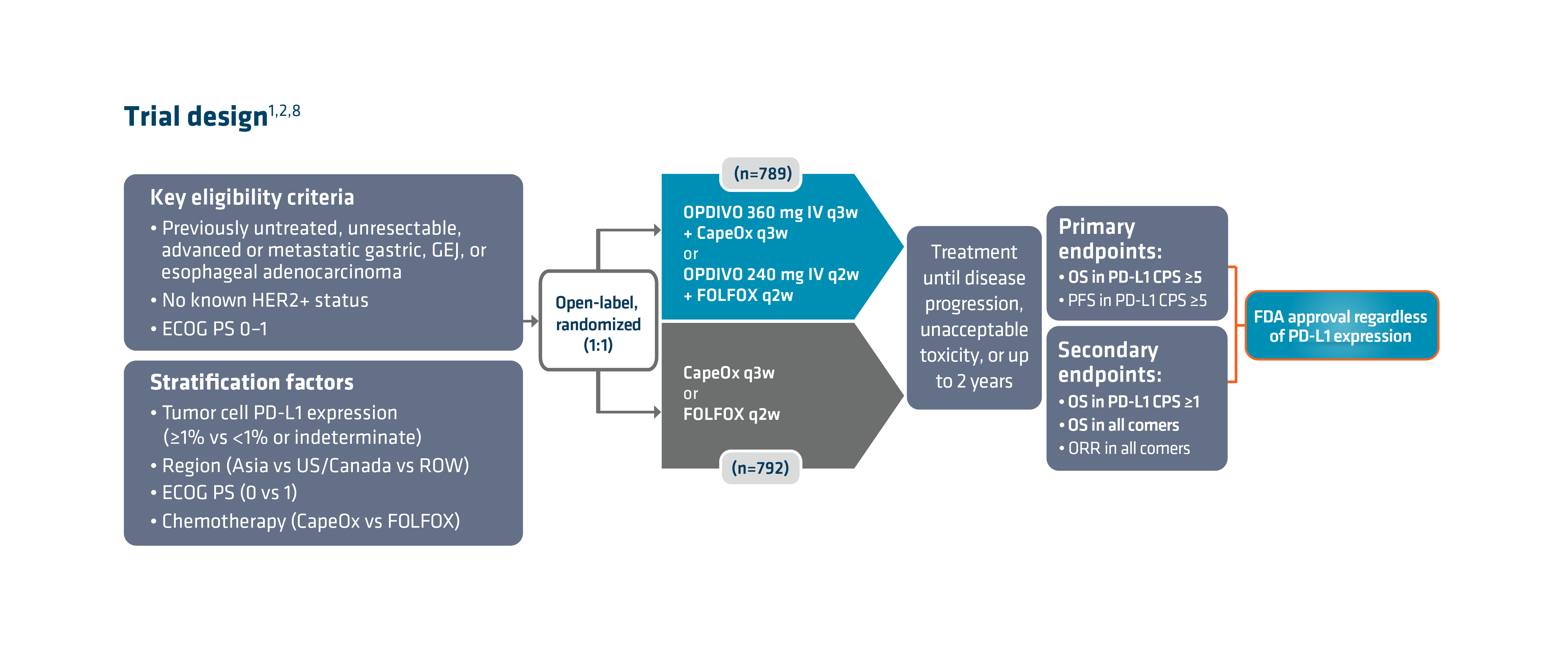
Trial-DesignCheckmate 649 enrolled 1581 patients randomized 1:1 to receive either OPDIVO + chemotherapy (n=789) or chemotherapy alone (n=792). The dual primary endpoints were OS and PFS in PD-L1 CPS ≥5. OS in PD-L1 CPS ≥1 and in all-comers were secondary endpoints, but were powered to measure statistical significance through hierarchical analysis. Baseline characteristics were consistent among all randomized patients and patients with PD-L1 CPS ≥5. Checkmate 649 was the first phase 3 trial to achieve positive results in the evaluation of a PD-1 inhibitor in combination with FOLFOX or CapeOx, allowing for synchronized I-O dosing options with the preferred chemotherapy.8

There are warnings and precautions associated with OPDIVO to keep in mind. These include severe and fatal immune-mediated adverse reactions, infusion-related reactions, complications of allogeneic hematopoietic stem cell transplantation, embryo-fetal toxicity, and increased mortality in patients with multiple myeloma when OPDIVO is added to a thalidomide analogue and dexamethasone, which is not recommended outside of controlled clinical trials.1 Additional information related to warnings and precautions can be found here .
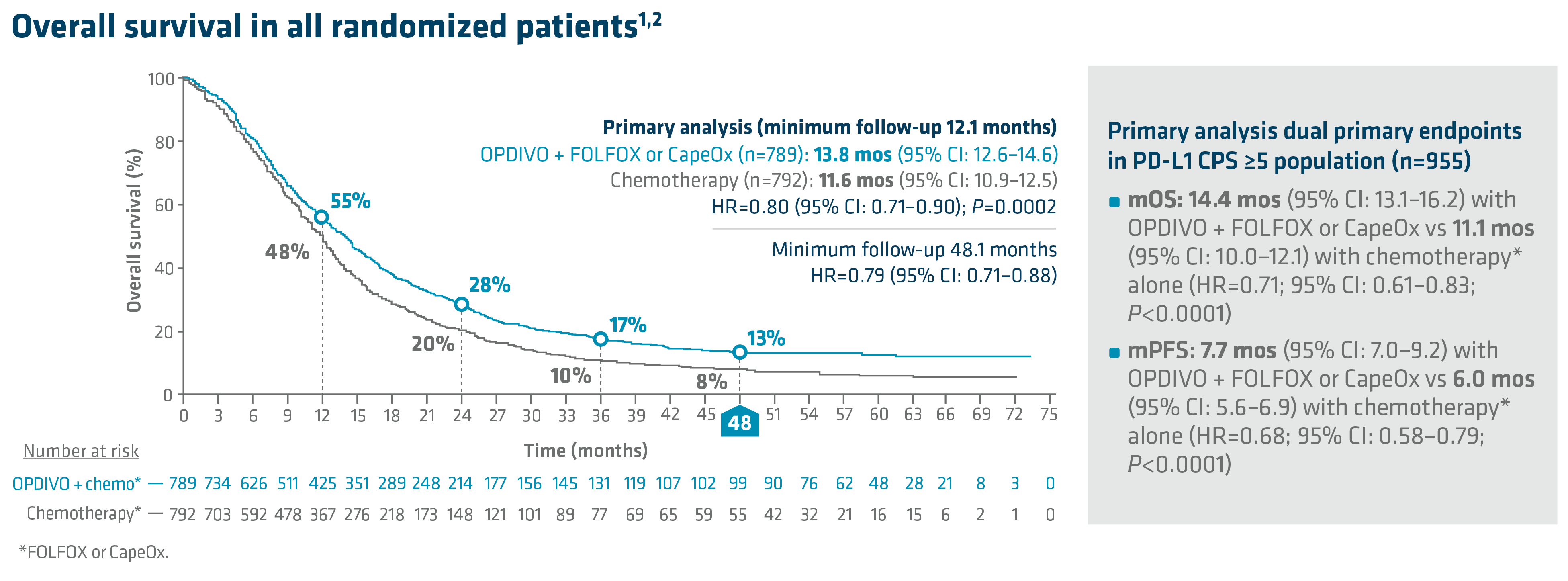
Overall-Survival-in-all-randomized-patientsIn the primary analysis (minimum follow-up of 12.1 months), OPDIVO + chemotherapy demonstrated superior OS in all randomized patients and patients with PD-L1 CPS ≥5, as compared to chemotherapy alone. In all randomized patients, mOS was 13.8 mos with OPDIVO + chemotherapy vs 11.6 mos with chemotherapy (HR=0.80; 95% CI: 0.71–0.90; P=0.0002). In patients with PD-L1 CPS ≥5 (n=955), mOS was 14.4 mos with OPDIVO + chemotherapy vs 11.1 mos with chemotherapy (HR=0.71; 95% CI: 0.61–0.83; P<0.0001).1 The 12-month OS rate in all randomized patients was 55% with OPDIVO + chemotherapy vs 48% with chemotherapy.8 “In my opinion, clinical trial data with OPDIVO + chemotherapy was a landmark. For the first time in a non–HER2-positive population, patients were able to break through the 1-year mOS barrier,” explained Dr. Kelly.

Durable survival data was observed for this OPDIVO-based regimen vs chemotherapy alone in GC/GEJC/EAC. The follow-up analysis at 48.1 months reported a mOS of 13.7 mos (95% CI: 12.4–14.5) with OPDIVO + chemotherapy vs 11.6 mos (95% CI: 10.9–12.5) with chemotherapy in all randomized patients (HR=0.79; 95% CI: 0.71–0.88), and 14.4 mos (95% CI: 13.1–16.2) with OPDIVO + chemotherapy vs 11.1 mos (95% CI: 10.1–12.1) with chemotherapy in patients with PD-L1 CPS ≥5 (HR=0.70; 95% CI: 0.61–0.81). The 48-month OS rate was 13% vs 8% for OPDIVO + chemotherapy vs chemotherapy, respectively, in all randomized patients.2

In Checkmate 649, the most common adverse reactions reported in ≥20% of patients treated with OPDIVO in combination with chemotherapy were peripheral neuropathy, nausea, fatigue, diarrhea, vomiting, decreased appetite, abdominal pain, constipation, and musculoskeletal pain. OPDIVO and/or chemotherapy were discontinued in 44% of patients and at least one dose was withheld in 76% of patients due to an adverse reaction. Serious adverse reactions occurred in 52% of patients treated with OPDIVO in combination with chemotherapy. The most frequent serious adverse reactions reported in ≥2% of patients treated with OPDIVO in combination with chemotherapy were vomiting (3.7%), pneumonia (3.6%), anemia (3.6%), pyrexia (2.8%), diarrhea (2.7%), febrile neutropenia (2.6%), and pneumonitis (2.4%). Fatal adverse reactions occurred in 16 (2.0%) patients who were treated with OPDIVO in combination with chemotherapy; these included pneumonitis (4 patients), febrile neutropenia (2 patients), stroke (2 patients), gastrointestinal toxicity, intestinal mucositis, septic shock, pneumonia, infection, gastrointestinal bleeding, mesenteric vessel thrombosis, and disseminated intravascular coagulation.1

An additional characteristic of OPDIVO is its flexible dosing schedule. Based on both the FDA-approved label and Checkmate 649 trial design, OPDIVO offers flexible synchronized dosing options based on chemotherapy preference, and “in my experience, allows scheduling according to the patient and clinician preference,” stated Dr. Kelly. Checkmate 649 evaluated OPDIVO (q2w or q3w) in combination with physician’s choice of either FOLFOX given q2w or CapeOx given q3w in the first-line treatment of certain metastatic gastroesophageal cancers. Treatment can be continued until disease progression, unacceptable toxicity, or up to 2 years.1
Synchronized-dosing-options-for-checkmate-649
Summary and conclusions

With the longest follow-up survival data in GC vs chemotherapy for any I-O–based regimen and durable survival data in GC/GEJC/EAC, OPDIVO in combination with fluoropyrimidine- and platinum-containing chemotherapy is an approved 1L treatment option for all eligible patients with non–HER2-positive GC/GEJC/EAC, regardless of PD-L1 status.1,2 OPDIVO also offers synchronized dosing options to align with preferred chemotherapies, including both FOLFOX and CapeOx, which can be used every 2 or 3 weeks, respectively.1 “I believe Checkmate 649 may act as a new benchmark moving forward and novel therapeutics may be compared against it,” stated Dr. Kelly.

1L=first line; CapeOx=capecitabine and oxaliplatin; CI=confidence interval; CNS=central nervous system; CPS=combined positive score; FOLFOX=leucovorin, fluorouracil, and oxaliplatin; GEJ=gastroesophageal junction; HER2=human epidermal growth factor receptor 2; HR=hazard ratio; I-O=immuno-oncology; IV=intravenous; mo=month; mOS=median OS; mPFS=median PFS; ORR=overall response rate; OS=overall survival; PD-1=programmed death receptor-1; PD-L1=programmed death ligand 1; PFS=progression-free survival; q2w=every 2 weeks; q3w=every 3 weeks; ROW=rest of world.

INDICATION
OPDIVO® (nivolumab), in combination with fluoropyrimidine- and platinum-containing chemotherapy, is indicated for the treatment of adult patients with advanced or metastatic gastric cancer, gastroesophageal junction cancer, and esophageal adenocarcinoma.
OPDIVO (10 mg/mL) is an injection for intravenous use.

IMPORTANT SAFETY INFORMATION

Severe and Fatal Immune-Mediated Adverse Reactions
• Immune-mediated adverse reactions listed herein may not include all possible severe and fatal immune-mediated adverse reactions.
• Immune-mediated adverse reactions, which may be severe or fatal, can occur in any organ system or tissue. While immune-mediated adverse reactions usually manifest during treatment, they can also occur after discontinuation of OPDIVO. Early identification and management are essential to ensure safe use of OPDIVO. Monitor for signs and symptoms that may be clinical manifestations of underlying immune-mediated adverse reactions. Evaluate clinical chemistries including liver enzymes, creatinine, and thyroid function at baseline and periodically during treatment with OPDIVO. In cases of suspected immune-mediated adverse reactions, initiate appropriate workup to exclude alternative etiologies, including infection. Institute medical management promptly, including specialty consultation as appropriate.
• Withhold or permanently discontinue OPDIVO depending on severity (please see section 2 Dosage and Administration in the accompanying Full Prescribing Information). In general, if OPDIVO interruption or discontinuation is required, administer systemic corticosteroid therapy (1 to 2 mg/kg/day prednisone or equivalent) until improvement to Grade 1 or less. Upon improvement to Grade 1 or less, initiate corticosteroid taper and continue to taper over at least 1 month. Consider administration of other systemic immunosuppressants in patients whose immune-mediated adverse reactions are not controlled with corticosteroid therapy. Toxicity management guidelines for adverse reactions that do not necessarily require systemic steroids (e.g., endocrinopathies and dermatologic reactions) are discussed below.
Immune-Mediated Pneumonitis
• OPDIVO can cause immune-mediated pneumonitis. The incidence of pneumonitis is higher in patients who have received prior thoracic radiation. In patients receiving OPDIVO monotherapy, immune-mediated pneumonitis occurred in 3.1% (61/1994) of patients, including Grade 4 (<0.1%), Grade 3 (0.9%), and Grade 2 (2.1%).
Immune-Mediated Colitis
• OPDIVO can cause immune-mediated colitis. A common symptom included in the definition of colitis was diarrhea. Cytomegalovirus (CMV) infection/reactivation has been reported in patients with corticosteroid-refractory immune-mediated colitis. In cases of corticosteroid-refractory colitis, consider repeating infectious workup to exclude alternative etiologies. In patients receiving OPDIVO monotherapy, immune-mediated colitis occurred in 2.9% (58/1994) of patients, including Grade 3 (1.7%) and Grade 2 (1%).
Immune-Mediated Hepatitis and Hepatotoxicity
• OPDIVO can cause immune-mediated hepatitis. In patients receiving OPDIVO monotherapy, immune-mediated hepatitis occurred in 1.8% (35/1994) of patients, including Grade 4 (0.2%), Grade 3 (1.3%), and Grade 2 (0.4%).
Immune-Mediated Endocrinopathies
• OPDIVO can cause primary or secondary adrenal insufficiency, immune-mediated hypophysitis, immune-mediated thyroid disorders, and Type 1 diabetes mellitus, which can present with diabetic ketoacidosis. Withhold OPDIVO depending on severity (please see section 2 Dosage and Administration in the accompanying Full Prescribing Information). For Grade 2 or higher adrenal insufficiency, initiate symptomatic treatment, including hormone replacement as clinically indicated. Hypophysitis can present with acute symptoms associated with mass effect such as headache, photophobia, or visual field defects. Hypophysitis can cause hypopituitarism; initiate hormone replacement as clinically indicated. Thyroiditis can present with or without endocrinopathy. Hypothyroidism can follow hyperthyroidism; initiate hormone replacement or medical management as clinically indicated. Monitor patients for hyperglycemia or other signs and symptoms of diabetes; initiate treatment with insulin as clinically indicated.
• In patients receiving OPDIVO monotherapy, adrenal insufficiency occurred in 1% (20/1994), including Grade 3 (0.4%) and Grade 2 (0.6%).
• In patients receiving OPDIVO monotherapy, hypophysitis occurred in 0.6% (12/1994) of patients, including Grade 3 (0.2%) and Grade 2 (0.3%).
• In patients receiving OPDIVO monotherapy, thyroiditis occurred in 0.6% (12/1994) of patients, including Grade 2 (0.2%).
• In patients receiving OPDIVO monotherapy, hyperthyroidism occurred in 2.7% (54/1994) of patients, including Grade 3 (<0.1%) and Grade 2 (1.2%).
• In patients receiving OPDIVO monotherapy, hypothyroidism occurred in 8% (163/1994) of patients, including Grade 3 (0.2%) and Grade 2 (4.8%).
• In patients receiving OPDIVO monotherapy, diabetes occurred in 0.9% (17/1994) of patients, including Grade 3 (0.4%) and Grade 2 (0.3%), and 2 cases of diabetic ketoacidosis.
Immune-Mediated Nephritis with Renal Dysfunction
• OPDIVO can cause immune-mediated nephritis. In patients receiving OPDIVO monotherapy, immune-mediated nephritis and renal dysfunction occurred in 1.2% (23/1994) of patients, including Grade 4 (<0.1%), Grade 3 (0.5%), and Grade 2 (0.6%).
Immune-Mediated Dermatologic Adverse Reactions
• OPDIVO can cause immune-mediated rash or dermatitis. Exfoliative dermatitis, including Stevens-Johnson syndrome (SJS), toxic epidermal necrolysis (TEN), and drug rash with eosinophilia and systemic symptoms (DRESS) has occurred with PD-1/PD-L1 blocking antibodies. Topical emollients and/or topical corticosteroids may be adequate to treat mild to moderate nonexfoliative rashes.
• Withhold or permanently discontinue OPDIVO depending on severity (please see section 2 Dosage and Administration in the accompanying Full Prescribing Information).
• In patients receiving OPDIVO monotherapy, immune-mediated rash occurred in 9% (171/1994) of patients, including Grade 3 (1.1%) and Grade 2 (2.2%).
Other Immune-Mediated Adverse Reactions
• The following clinically significant immune-mediated adverse reactions occurred at an incidence of <1% (unless otherwise noted) in patients who received OPDIVO monotherapy or were reported with the use of other PD-1/PD-L1 blocking antibodies. Severe or fatal cases have been reported for some of these adverse reactions: cardiac/vascular: myocarditis, pericarditis, vasculitis; nervous system: meningitis, encephalitis, myelitis and demyelination, myasthenic syndrome/myasthenia gravis (including exacerbation), Guillain-Barré syndrome, nerve paresis, autoimmune neuropathy; ocular: uveitis, iritis, and other ocular inflammatory toxicities can occur; gastrointestinal: pancreatitis to include increases in serum amylase and lipase levels, gastritis, duodenitis; musculoskeletal and connective tissue: myositis/polymyositis, rhabdomyolysis, and associated sequelae including renal failure, arthritis, polymyalgia rheumatica; endocrine: hypoparathyroidism; other (hematologic/immune): hemolytic anemia, aplastic anemia, hemophagocytic lymphohistiocytosis (HLH), systemic inflammatory response syndrome, histiocytic necrotizing lymphadenitis (Kikuchi lymphadenitis), sarcoidosis, immune thrombocytopenic purpura, solid organ transplant rejection.
• Some ocular IMAR cases can be associated with retinal detachment. Various grades of visual impairment, including blindness, can occur. If uveitis occurs in combination with other immune-mediated adverse reactions, consider a Vogt-Koyanagi-Harada–like syndrome, which has been observed in patients receiving OPDIVO, as this may require treatment with systemic corticosteroids to reduce the risk of permanent vision loss.
Infusion-Related Reactions
• OPDIVO can cause severe infusion-related reactions. Discontinue OPDIVO in patients with severe (Grade 3) or life-threatening (Grade 4) infusion-related reactions. Interrupt or slow the rate of infusion in patients with mild (Grade 1) or moderate (Grade 2) infusion-related reactions. In patients receiving OPDIVO monotherapy as a 60-minute infusion, infusion-related reactions occurred in 6.4% (127/1994) of patients. In a separate trial in which patients received OPDIVO monotherapy as a 60-minute infusion or a 30-minute infusion, infusion-related reactions occurred in 2.2% (8/368) and 2.7% (10/369) of patients, respectively. Additionally, 0.5% (2/368) and 1.4% (5/369) of patients, respectively, experienced adverse reactions within 48 hours of infusion that led to dose delay, permanent discontinuation or withholding of OPDIVO.
Complications of Allogeneic Hematopoietic Stem Cell Transplantation
• Fatal and other serious complications can occur in patients who receive allogeneic hematopoietic stem cell transplantation (HSCT) before or after being treated with OPDIVO. Transplant-related complications include hyperacute graft-versus-host-disease (GVHD), acute GVHD, chronic GVHD, hepatic veno-occlusive disease (VOD) after reduced intensity conditioning, and steroid-requiring febrile syndrome (without an identified infectious cause). These complications may occur despite intervening therapy between OPDIVO and allogeneic HSCT.
• Follow patients closely for evidence of transplant-related complications and intervene promptly. Consider the benefit versus risks of treatment with OPDIVO prior to or after an allogeneic HSCT.
Embryo-Fetal Toxicity
• Based on its mechanism of action and findings from animal studies, OPDIVO can cause fetal harm when administered to a pregnant woman. Advise pregnant women of the potential risk to a fetus. Advise females of reproductive potential to use effective contraception during treatment with OPDIVO and for at least 5 months after the last dose.
Increased Mortality in Patients with Multiple Myeloma when OPDIVO is Added to a Thalidomide Analogue and Dexamethasone
• In randomized clinical trials in patients with multiple myeloma, the addition of OPDIVO to a thalidomide analogue plus dexamethasone resulted in increased mortality. Treatment of patients with multiple myeloma with a PD-1 or PD-L1 blocking antibody in combination with a thalidomide analogue plus dexamethasone is not recommended outside of controlled clinical trials.
Lactation
• There are no data on the presence of OPDIVO in human milk, the effects on the breastfed child, or the effects on milk production. Because of the potential for serious adverse reactions in breastfed children, advise women not to breastfeed during treatment and for 5 months after the last dose.
Serious Adverse Reactions
• In Checkmate 649, serious adverse reactions occurred in 52% of patients treated with OPDIVO in combination with chemotherapy (n=782). The most frequent serious adverse reactions reported in ≥2% of patients treated with OPDIVO in combination with chemotherapy were vomiting (3.7%), pneumonia (3.6%), anemia (3.6%), pyrexia (2.8%), diarrhea (2.7%), febrile neutropenia (2.6%), and pneumonitis (2.4%). Fatal adverse reactions occurred in 16 (2.0%) patients who were treated with OPDIVO in combination with chemotherapy; these included pneumonitis (4 patients), febrile neutropenia (2 patients), stroke (2 patients), gastrointestinal toxicity, intestinal mucositis, septic shock, pneumonia, infection, gastrointestinal bleeding, mesenteric vessel thrombosis, and disseminated intravascular coagulation.
Common Adverse Reactions
• In Checkmate 649, the most common adverse reactions (≥20%) in patients treated with OPDIVO in combination with chemotherapy (n=782) were peripheral neuropathy (53%), nausea (48%), fatigue (44%), diarrhea (39%), vomiting (31%), decreased appetite (29%), abdominal pain (27%), constipation (25%), and musculoskeletal pain (20%).

Please see US Full Prescribing Information for OPDIVO.

References:

1. OPDIVO [package insert]. Princeton, NJ: Bristol-Myers Squibb Company.
2. Shitara K, Moehler M, Ajani JA, et al. Nivolumab plus chemotherapy vs chemotherapy as first-line treatment for advanced gastric cancer/gastroesophageal junction cancer/esophageal adenocarcinoma: 4-year follow-up of the CheckMate 649 study. Oral presentation at ASCO GI 2024. Abstract 306.
3. Mantziari S, St Amour P, Abboretti F, et al. A comprehensive review of prognostic factors in patients with gastric adenocarcinoma. Cancers (Basel). 2023;15(5):1628.
4. Imamura Y, Watanabe M, Oki E, Morita M, Baba H. Esophagogastric junction adenocarcinoma shares characteristics with gastric adenocarcinoma: literature review and retrospective multicenter cohort study. Ann Gastroenterol Surg. 2020;5(1):46-59.
5. Rogers MP, DeSantis AJ, DuCoin CG. Oligometastatic adenocarcinoma of the esophagus: current understanding, diagnosis, and therapeutic strategies. Cancers (Basel). 2021;13(17):4352.
6. Paydary K, Reizine N, Catenacci DVT. Immune-checkpoint inhibition in the treatment of gastro-esophageal cancer: a closer look at the emerging evidence. Cancers (Basel). 2021;13(23):5929.
7. Sung H, Ferlay J, Siegel RL, et al. Global cancer statistics 2020: GLOBOCAN estimates of incidence and mortality worldwide for 36 cancers in 185 countries. CA Cancer J Clin. 2021;71(3):209-249.
8. Janjigian YY, Shitara K, Moehler M, et al. First-line nivolumab plus chemotherapy versus chemotherapy alone for advanced gastric, gastro-oesophageal junction, and oesophageal adenocarcinoma (CheckMate 649): a randomised, open-label, phase 3 trial. Lancet. 2021;398(10294):27-40.
9. Janjigian YY, Ajani JA, Moehler M, et al. Nivolumab plus chemotherapy or ipilimumab vs chemotherapy as first-line treatment for advanced gastric cancer/gastroesophageal junction cancer/esophageal adenocarcinoma: CheckMate 649 study. Oral presentation at ESMO 2021. Abstract LBA7.
10. Shankaran V, Xiao H, Bertwistle D, et al. A comparison of real-world treatment patterns and clinical outcomes in patients receiving first-line therapy for unresectable advanced gastric or gastroesophageal junction cancer versus esophageal adenocarcinomas. Adv Ther. 2021;38:
707-720.
11. BMS-REF-NIVO-0256. Princeton, NJ: Bristol-Myers Squibb Company; 2024.

© 2024 Bristol-Myers Squibb Company. OPDIVO® is a registered trademark of Bristol-Myers Squibb Company.
1506-US-2300555 01/24

New ASCO Guideline Recommends Germline Testing in ALL Newly Diagnosed Breast Cancer Patients 65 Years or Younger

SUMMARY: Breast cancer is the most common cancer among women in the US and about 1 in 8 women (12%) will develop invasive breast cancer during their lifetime. It is estimated that in the US, approximately 310,720 new cases of female breast cancer will be diagnosed in 2024, and about 42,250 individuals will die of the disease, largely due to metastatic recurrence.

The availability of multigene panel testing and next-generation sequencing can change the landscape of cancer prevention and treatment. However, there is lack of guidance for clinicians on whom to test and/or which genes to include in germline genetic testing panels for Pathogenic Variants.

The American Society of Clinical Oncology along with the Society of Surgical Oncology on January 4, 2024 provided new clinical practice guideline for clinicians and other Health Care Providers, regarding the role of germline mutation testing in patients with breast cancer, based on the best available evidence. These recommendations were developed based on a systematic review of 47 articles that met eligibility criteria for the germline mutation testing recommendations, and 18 articles that met eligibility criteria for the genetic counseling recommendations.

The guideline addressed the following question: Which patients with breast cancer should have germline genetic testing for Pathogenic Variants (PVs) in cancer susceptibility genes?

Question 1. Should clinicians offer BRCA1/2 testing to all patients with newly diagnosed breast cancer?
Recommendation 1.1
All patients newly diagnosed with breast cancer with Stage I-III or de novo Stage IV/metastatic disease who are 65 years or younger at diagnosis should be offered BRCA1/2 testing.
Recommendation 1.2
All patients newly diagnosed with breast cancer with Stage I-III or de novo Stage IV/metastatic disease who are older than age 65 should be offered BRCA1/2 testing if:
a) They are candidates for poly(ADP–ribose) polymerase (PARP) inhibitor therapy for early-stage or metastatic disease.
b) They have triple-negative breast cancer.
c) Their personal or family history suggests the possibility of a pathogenic variant.
d) They were assigned male sex at birth.
e) They are of Ashkenazi Jewish ancestry or are members of a population with an increased prevalence of founder mutations.
Recommendation 1.3
Patients undergoing BRCA1/2 testing should also be offered testing for other cancer predisposition genes as suggested by their personal or family history. Consultation with a provider experienced in clinical cancer genetics can help guide this decision-making and should be made available to patients when possible.

Question 2. Should all people with recurrent disease, local or metastatic, or with second breast primary, be offered BRCA1/2 testing?
Recommendation 2.1
All patients with recurrent breast cancer (local or metastatic) who are candidates for PARP inhibitor therapy should be offered BRCA1/2 testing regardless of family history.
Qualifying statement.
Small single-arm studies show that oral PARP inhibitor therapy demonstrates high response rates in women with metastatic breast cancer and germline pathogenic variants in PALB2.
Recommendation 2.2
BRCA1/2 testing should be offered to patients with a second primary cancer either in the contralateral or ipsilateral breast.

Question 3. Should people with a personal history of breast cancer (and no active disease) be offered BRCA1/2 testing?
Recommendation 3.1
All patients with a personal history of breast cancer diagnosed 65 years or less who are without active disease should be offered BRCA1/2 testing if the result will inform personal risk management or family risk assessment.
Recommendation 3.2
All patients with a personal history of breast cancer diagnosed over age 65 with no active disease, who meet one of the following criteria, should be offered BRCA1/2 testing if the result will inform personal risk management or family risk assessment:
a) Their personal or family history suggests the possibility of a pathogenic variant.
b) They were assigned male sex at birth.
c) They had triple-negative breast cancer.
d) They are of Ashkenazi Jewish ancestry or are members of a population with an increased prevalence of founder mutations.

Question 4. What is the value of testing patients with a diagnosis of breast cancer for breast cancer predisposition genes other than BRCA1/2?
Recommendation 4.1
Testing for high penetrance genes beyond BRCA1/2, including PALB2, TP53, PTEN, STK11, and CDH1, could inform medical therapy, influence surgical decision making, refine estimates of risks of second primary cancer, and inform family risk assessment, and thus should be offered to appropriate patients.
Recommendation 4.2
Testing for moderate penetrance breast cancer genes currently offers no benefits for treatment of the index breast cancer but may inform risks of second primary cancer or family risk assessment, and thus may be offered to appropriate patients who are undergoing BRCA1/2 testing.
Recommendation 4.3
If a multi-gene panel is ordered, the specific panel chosen should take into account the patients personal and family history. Consultation with a provider experienced in clinical cancer genetics can be helpful in selecting a specific multi-gene panel or interpreting its results and should be made available to patients when possible.

Question 5. How should patients with breast cancer considering genetic testing be counseled?
Recommendation 5.1
Patients undergoing genetic testing should be given sufficient information before testing to provide informed consent.
Recommendation 5.2
Patients with pathogenic variants should be provided with individualized post-test genetic counseling and offered referral to a provider experienced in clinical cancer genetics.
Recommendation 5.3
Variants of uncertain significance should not alter management. Patients should be made aware that variants of uncertain significance may be reclassified as being pathogenic, and they should understand that periodic follow up is necessary. Consultation with a provider experienced in clinical cancer genetics can be helpful and should be made available to patients when possible.
Recommendation 5.4
Patients without a pathogenic variant on genetic testing may still benefit from counseling, if there is a significant family history of cancer, and referral to a provider experienced in clinical cancer genetics is recommended.

ASCO believes that cancer clinical trials are vital to inform medical decisions and improve cancer care, and that all patients should have the opportunity to participate.

Germline Testing in Patients With Breast Cancer: ASCO–Society of Surgical Oncology Guideline. Bedrosian I, MD , Somerfield MR, PhD, Achatz MI, et al. Journal of Clinical Oncology January 04, 2024. https://doi.org/10.1200/JCO.23.02225.

Late Breaking Abstract – ASH 2023: Oral Ibrutinib-Venetoclax Combination Improved Outcomes in Mantle Cell Lymphoma

SUMMARY: It is estimated that approximately 3,300 new cases of Mantle Cell Lymphoma (MCL) are diagnosed in the US each year. Mantle Cell Lymphoma is an aggressive B-cell lymphoma and accounts for approximately 6% of all Non Hodgkin Lymphomas in adults, and is associated with a high relapse rate following dose-intensive therapies. Early and late relapses in patients with MCL have been attributed to persistence of residual disease.

Majority of patients with MCL are elderly and are not candidates for aggressive treatment or Autologous Stem Cell Transplantation. The four BTK inhibitors presently approved by the FDA for MCL include IMBRUVICA® (Ibrutinib) approved in 2013, CALQUENCE® (Acalabrutinib) approved in 2017, BRUKINSA® (Zanubrutinib) approved in 2019 and JAYPIRCA® (Pirtobrutinib) approved in 2023.

Brutons Tyrosine Kinase (BTK) is a member of the Tec family of kinases, downstream of the B-cell receptor, and is predominantly expressed in B-cells. It is a mediator of B-cell receptor signaling in normal and transformed B-cells. BTK inhibitors inhibit cell proliferation, and promote programmed cell death (Apoptosis) by blocking B-cell activation and signaling. Single agent Ibrutinib is presently approved by the FDA for the treatment of MCL patients who have received at least one prior therapy. Venetoclax (VENCLEXTA®) is a second generation, oral, selective, small molecule inhibitor of BCL2 and restores the apoptotic processes in tumor cells. By virtue of their unique and complementary mechanism of action, Ibrutinib in combination with Venetoclax demonstrated promising clinical activity in early phase MCL studies (N Engl J Med 2018; 378:1211-1223).

The Sympatico trial, is a multinational, randomized, double-blind, phase III study conducted to compare the combination of Ibrutinib and Venetoclax with Ibrutinib plus placebo, in patients with relapsed or refractory Mantle Cell Lymphoma (MCL). In this study, a total of 267 adult patients (N=267) with relapsed or refractory MCL who had previously received at least one prior line of therapy were randomly assigned in a 1:1 ratio to receive Ibrutinib 560 mg orally once daily concurrently with, either oral Venetoclax given at a standard 5-week ramp-up dose to a target dose of 400 mg once daily (N=134), or Placebo (N=133) for 2 years, followed by single-agent Ibrutinib until progressive disease (PD) or unacceptable toxicity. The median age was 68 years, 96% of patients had an ECOG PS of 0-1, 17% had 3 or more prior lines of therapy, and 22% were at increased risk for Tumor Lysis Syndrome (TLS). Both treatment groups were well balanced, and randomization was stratified based on ECOG PS, prior lines of therapy, and TLS risk based on tumor burden and Creatinine Clearance. The study evaluated the efficacy of the combination therapy across various subgroups, including those with high-risk features such as blastoid variant or TP53-mutated MCL. The Primary endpoint was investigator assessed Progression Free Survival (PFS) using Lugano criteria, and key Secondary endpoints included Complete Response (CR) rate, Time To Next Treatment (TTNT), Overall Survival (OS), and Overall Response Rate (ORR) by investigator assessment.

With a median follow up of 51.2 months, the median PFS was significantly longer with the Ibrutinib-Venetoclax combination, compared with the Placebo group (31.9 months versus 22.1 months; HR=0.65; P=0.0052). These PFS benefits were consistent across patient subgroups, including those with blastoid-variant or TP53-mutated MCL. In the combination group, 54% of patients achieved a Complete Remission, compared to 32% in the Placebo group (P=0.0004). The Time to Next Treatment in the combination group was median Not Reached (NR) versus 35.4 months in the Placebo group. At the time of this primary analysis, the median OS was 44.9 months with the Ibrutinib-Venetoclax combination versus 38.6 months with Ibrutinib plus Placebo, but the difference was not statistically significant. Adverse events were more common among patients who received the combination therapy, and included cytopenias and pneumonia.

It was concluded that a combination of Ibrutinib and Venetoclax was synergistic and demonstrated efficacy and safety, for the treatment of relapsed or refractory Mantle Cell Lymphoma, providing a potential new standard of care for this patient population. This chemo-free treatment option represents a milestone achievement in Mantle Cell Lymphoma treatment.

Ibrutinib Combined with Venetoclax in Patients with Relapsed/Refractory Mantle Cell Lymphoma: Primary Analysis Results from the Randomized Phase 3 Sympatico Study. Wang M, Jurczak W, Trněný M, et al. Presented at the 2023 ASH Annual Meeting & Exposition December 9-12, 2023. LBA-2.

Neoadjuvant Chemoimmunotherapy Improves Pathologic Complete Response Rates in Early Stage ER-Positive, HER2-Negative Breast Cancer

SUMMARY: Breast cancer is the most common cancer among women in the US and about 1 in 8 women (12%) will develop invasive breast cancer during their lifetime. It is estimated that in the US, approximately 310,720 new cases of female breast cancer will be diagnosed in 2024, and about 42,250 individuals will die of the disease, largely due to metastatic recurrence. About 70% of breast tumors express Estrogen Receptors and/or Progesterone Receptors, and Hormone Receptor (HR)-positive/HER2-negative breast cancer is the most frequently diagnosed molecular subtype. Approximately 25% of patients with early-stage breast cancer who receive (neo)adjuvant chemotherapy experience a recurrence within 5 years (Lancet Oncol. 2018;19:27-39).

The promising efficacy observed with single-agent checkpoint blockade for advanced HER2-negative breast cancer, and the significant benefit observed with PD-1 inhibitors combined with chemotherapy for lung cancer and other cancer types, led the researchers to evaluate the efficacy of adding Pembrolizumab to standard neoadjuvant chemotherapy. In the Phase 2 I-SPY2 trial, Pembrolizumab plus neoadjuvant chemotherapy improved estimated pathological Complete Response rates versus neoadjuvant chemotherapy alone, at 30% versus 13%, in patients with HR-positive, HER2-negative breast cancer.

Pembrolizumab (KEYTRUDA®) is a fully humanized, Immunoglobulin G4, anti-PD-1, monoclonal antibody, that binds to the PD-1 receptor and blocks its interaction with ligands PD-L1 and PD-L2. It thereby reverses the PD-1 pathway-mediated inhibition of the immune response and unleashes the tumor-specific effector T cells. The rationale for combining chemotherapy with immunotherapy is that cytotoxic chemotherapy releases tumor-specific antigens, and immune checkpoint inhibitors such as Pembrolizumab when given along with chemotherapy can enhance endogenous anticancer immunity.

Pembrolizumab is approved for the treatment of patients with high-risk early-stage Triple Negative Breast Cancer (TNBC) in combination with chemotherapy as neoadjuvant treatment, as well as in combination with chemotherapy for the treatment of patients with locally recurrent unresectable or metastatic TNBC whose tumors express PD-L1 (CPS 10 or more).

KEYNOTE-756 is a global, randomized, double-blind, Phase III trial, conducted to assess the efficacy and safety of Pembrolizumab versus placebo, in combination with neoadjuvant chemotherapy followed by adjuvant treatment with Pembrolizumab plus endocrine therapy, in adults with high-risk, early stage ER-positive HER2- negative breast cancer. In this study 1,278 enrolled patients (N=1278) were randomized 1:1 to receive Pembrolizumab 200 mg IV ever 3 weeks or placebo, both given with Paclitaxel weekly for 12 weeks, followed by 4 additional cycles of Doxorubicin or Epirubicin plus Cyclophosphamide (neoadjuvant treatment) prior to surgery. Following definitive surgery with or without radiation treatment, patients received Pembrolizumab or placebo every 3 weeks for 9 cycles plus endocrine therapy for up to 10 years, as adjuvant therapy post-surgery. Eligible patients had centrally confirmed T1c-2 (≥2 cm) cN1-2 or T3-4 cN0-2, Grade 3, ER-positive, HER2-negative, invasive ductal carcinoma, and were treatment-naive. Both treatment groups were well balanced. The median age was 49 years, about 76% of patients in each treatment group had a PD-L1 CPS of 1 or higher, about 40% had a CPS of 10 or higher, and about 90% had nodal involvement. About 62% of patients had Stage II disease, and 38% had Stage III disease. The dual Primary endpoints were pathological Complete Response (pCR) rate (ypT0/Tis ypN0), defined as absence of invasive cancer in the breast and axillary lymph nodes at the time of surgery, and Event Free Survival (EFS). Secondary endpoints included Overall Survival and Safety.

With a median follow-up of 33.2 months, the study demonstrated a statistically significant improvement in pCR rates with Pembrolizumab compared to placebo. The pCR rate in the intention-to-treat (ITT) population was 24.3% with Pembrolizumab versus 15.6% with placebo (absolute difference 8.5%; P = 0.00005). Similar improvements were observed across various subgroups, including patients with Stage II or III disease, positive lymph nodes at baseline, and higher PD-L1 expression levels. Pembrolizumab demonstrated superior efficacy across geographic regions and exhibited a linear improvement in pCR rates with increasing PD-L1 expression.

Further analyses showed a greater pCR benefit with Pembrolizumab in patients with low estrogen receptor (ER) positivity (defined as less than 10% of ER-positive cells), node positive disease and those with higher PD-L1 expression. Pembrolizumab recipients who received full-dose chemotherapy had a greater pCR benefit compared to those who received reduced chemotherapy doses. Additionally, Pembrolizumab recipients were more likely to shift to lower Residual Cancer Burden (RCB) groups post-surgery. The trial also observed higher rates of immune-mediated adverse events with Pembrolizumab compared to placebo, with common events including hypothyroidism, hyperthyroidism, and pneumonitis.

It was concluded from this study that, the addition of Pembrolizumab to neoadjuvant chemotherapy followed by adjuvant Pembrolizumab plus endocrine therapy, significantly improves pCR rates in patients with early stage, high risk ER-positive, HER2-negative breast cancer. Further assessment of long term outcomes, including Event-Free Survival and Overall Survival is ongoing to fully evaluate the clinical benefit of this treatment approach. The study sponsors added that this is the first positive Phase III study, evaluating an immunotherapy-based regimen for patients with high risk, early stage ER-positive, HER2-negative breast cancer, and an important milestone in advancing research, in early stage breast cancer.

Phase 3 study of neoadjuvant pembrolizumab or placebo plus chemotherapy, followed by adjuvant pembrolizumab or placebo plus endocrine therapy for early-stage high-risk ER+/HER2- breast cancer: KEYNOTE-756. Cardoso F, O’Shaughnessy J, McArthur H, et al. Presented at SABCS 2023. December 5-9, 2023. San Antonio, TX. Abstract GS01-02.

Late Breaking Abstract: ASH – 2023: Pomalidomide Reduces Epistaxis and Improves Quality of Life in Hereditary Hemorrhagic Telangiectasia

SUMMARY: Hereditary Hemorrhagic Telangiectasia (HHT) is an Autosomal Dominant inherited disorder caused by mutations in regulators of angiogenesis. Also known as Osler-Weber-Rendu syndrome, HHT is the second most common inherited bleeding disorder after Von Willebrand Disease, with an estimated prevalence of 1 in 5000. HHT presents with a triad of recurrent epistaxis with iron deficiency anemia, mucocutaneous telangiectasias, and visceral arteriovenous malformations (AVMs) and in more severe cases, patients may experience life-threatening hemorrhage, stroke, or high-output heart failure, requiring hospitalizations, with a negative impact on Quality of Life (QOL). HHT is caused by disruptions in angiogenesis signaling, resulting in impaired vascular development. Three genes in the Transforming Growth Factor-beta (TGF-β) signaling pathway have been implicated and they include Endoglin (ENG), activin A receptor ligand type I (ACVRL1 or ALK-1), and SMAD family member 4 (MADH4 or SMAD4).

Small non-randomized studies suggested that systemic antiangiogenic agent Bevacizumab or immunomodulatory drugs with antiangiogenic properties such as Thalidomide, Lenalidomide, and Pomalidomide may be effective in treating HHT. There are presently no FDA approved therapies for HHT.

PATH-HHT is a randomized, placebo-controlled, multicenter clinical trial, conducted in the US to determine the safety and efficacy of Pomalidomide, for bleeding in HHT. In this study, 144 patients (N=144) diagnosed with HHT were randomly assigned in a 2:1 ratio to receive either Pomalidomide 4 mg orally daily or a matching placebo, for a duration of six months. Pomalidomide, instead of another immunomodulatory drug, was chosen due to its favorable safety profile. Eligibility criteria included a confirmed HHT diagnosis per Curaçao Diagnostic Criteria, documented anemia, and an Epistaxis Severity Score (ESS) of 3 or more over the prior 3 months. Epistaxis Severity Score (ESS) was developed to self- describe epistaxis severity from 0-10, with 10 representing the most severe epistaxis. Mild is ESS of 1-4, moderate is ESS of 4-7 and Severe is ESS of 7-10. The mean age was 59 years and 48% were female. Among the 134 patients who agreed to genetic testing, ENG mutations were detected in 37%, ACVRL1 in 51%, and SMAD4 in 1%. Patients had a mean ESS of 5 at baseline, and mean daily epistaxis duration of 16 minutes. In the preceding 6 months, 84% of patients had required iron infusions and 19% required blood transfusions. More than a third of the patients also had GI bleeding, and 40% had pulmonary AVMs. The Primary endpoint of the study was the change in Epistaxis Severity Score (ESS), from baseline to the end of the six-month treatment period. Secondary endpoints included changes in the average daily self-reported duration of epistaxis from the 4 weeks preceding the baseline visit to weeks 20-24 of treatment, the amount of parenteral iron infused or blood transfused, and change in Quality-of-Life (QOL) measurements, including an HHT-specific QOL score.

The results of this study showed that treatment with Pomalidomide led to a significant reduction in epistaxis severity compared to placebo. The mean ESS decreased by -1.84 in the Pomalidomide group versus -0.89 in the placebo group at 24 weeks (P=0.003). This benefit was seen as early as week 12. Additionally, patients treated with Pomalidomide reported greater improvements in Quality of Life (QOL) related to HHT. The HHT-specific QOL score (ranges from 0-16 with higher scores indicating more limitations) also decreased more in the Pomalidomide group versus the placebo group at 24 weeks (P=0.015). Adverse events were more common in the Pomalidomide group and included mild to moderate neutropenia (45% versus 10%), constipation/diarrhea (60% versus 37%), and rash (36% versus 10%).

It was concluded from this largest HHT study that treatment with Pomalidomide demonstrated a significant and highly clinically relevant reduction in epistaxis, as well as an improvement in the HHT-specific QOL score. Pomalidomide holds promise as a therapeutic option for patients with HHT, addressing an unmet medical need, in managing this challenging genetic disorder. Additional studies may identify biomarkers predicting responses to Pomalidomide.

PATH-HHT, a Double-Blind, Randomized, Placebo-Controlled Trial in Hereditary Hemorrhagic Telangiectasia Demonstrates That Pomalidomide Reduces Epistaxis and Improves Quality of Life. Al-Samkari H, Kasthuri RS, Iyer V, et al. Blood (2023) 142 (Supplement 2): LBA-3. https://doi.org/10.1182/blood-2023-191983.

Dato-DXd for Patients with HR-Positive HER2-Negative Advanced Breast Cancer

SUMMARY: Breast cancer is the most common cancer among women in the US and about 1 in 8 women (12%) will develop invasive breast cancer during their lifetime. It is estimated that in the US, approximately 310,720 new cases of female breast cancer will be diagnosed in 2024, and about 42,250 individuals will die of the disease, largely due to metastatic recurrence.

Approximately 70% of breast tumors in patients with metastatic disease are Estrogen Receptor (ER) and/or Progesterone Receptor (PR) positive and HER2-negative. These patients are often treated with single agent endocrine therapy, endocrine therapy in combination with CDK4/6 inhibitor, or chemotherapy. Resistance to hormonal therapy occurs in a majority of the patients and there is therefore an unmet need for agents with novel mechanisms of action.

Datopotamab-deruxtecan (Dato-DXd) is an ADC composed of a TROP2-directed monoclonal antibody conjugated to a potent topoisomerase I inhibitor via a stable tetrapeptide-based cleavable linker. Trop-2 is a transmembrane calcium signal transducer that stimulates cancer cell growth. Trop-2 is overexpressed in several epithelial cancers including cancers of the breast, colon and lung, and has limited expression in normal human tissues. It has been associated with poor Overall and Disease-Free Survival in several types of solid tumors. Trop-2 is expressed in more than 85% of breast tumors including Triple Negative Breast Cancer. Upon binding to Trop-2, the anti-TROP-2 monoclonal antibody is internalized and delivers the payload directly into the tumor cell, making it a suitable transporter for the delivery of cytotoxic drugs. Further, the cleavable linker enables the payload to be released both intracellularly into the tumor cells, as well as the tumor microenvironment, thereby allowing for the delivery of therapeutic concentrations of the active drug in bystander cells to which the conjugate has not bound. Dato-DXd showed encouraging antitumor activity in the TROPION-PanTumor01 trial, an ongoing multicenter, open-label study, evaluating Dato-DXd in different dose levels in solid tumors.

TROPION-Breast01 is an open-label, global, Phase III study in which 732 patients (N=732) with HR-positive HER2-negative previously treated metastatic breast cancer were randomly assigned in a 1:1 manner to receive either Dato-DXd (N=365) or investigators choice of chemotherapy (N=367). Dato-DXd was given at a dose of 6 mg/kg IV on day 1 every 3 weeks. Investigators choice of chemotherapy consisted of Eribulin mesylate, Vinorelbine, or Gemcitabine, all given IV on days 1 and 8 every 3 weeks, as well as Capecitabine given orally on days 1-14 every 3 weeks. Treatment was continued until disease progression or unacceptable toxicities. The median age was 55 years and enrolled patients had received 1 or 2 prior lines of chemotherapy in the inoperable or metastatic setting. Eligible patients had progressed on, or were deemed unsuitable for endocrine therapy. Patients were stratified by number of lines of chemotherapy received in the unresectable/metastatic setting, and treatment with a previous CDK4/6 inhibitor. The Co-Primary end points were Progression Free Survival (PFS) by Blinded Independent Central Review (BICR) and Overall Survival (OS). Secondary end points included Overall Response Rate (ORR), Safety, Patient Reported Outcomes, and Time to First Subsequent Therapy (TFST).

The Median PFS by BICR in the Dato-DXd arm was 6.9 months versus 4.9 months in the chemotherapy arm (HR=0.63; P < 0.0001). The PFS rates at 6, 9, and 12-months in Dato-DXd arm were 55.2%, 34.7%, and 21.7%, respectively. In the chemotherapy arm, these rates were 36.9%, 20.9%, and 9.9%, respectively. The PFS benefit with Dato-DXd over chemotherapy was noted irrespective of brain metastases and prior duration of treatment with CDK4/6 inhibitors. The median Time to First Subsequent Therapy was 8.2 months with Dato-DXd and 5.0 months with chemotherapy. Dato-DXd also demonstrated a delay in time to deterioration in global health status/quality of life, compared to chemotherapy.

Treatment-related adverse effects occurred in 94% of patients in the Dato-DXd arm versus 86% in the chemotherapy group, with grade 3 or higher severity in 21% versus 45%, respectively. Neutropenia was more common in the chemotherapy arm.

It was concluded that Dato-DXd showed statistically significant and clinically meaningful improvement in Progression Free Survival compared to chemotherapy. The improved outcomes were observed across subgroups, including patients with and without brain metastases, and those with varying durations of prior CDK4/6 inhibitor treatment. Dato-DXd was associated with a favorable safety profile and impact on quality of life.

Randomized phase 3 study of datopotamab deruxtecan vs chemotherapy for patients with previously-treated inoperable or metastatic hormone receptor-positive, HER2-negative breast cancer: Results from TROPION-Breast01. Bardia A, Jhaveri K, Im S-A, et al. Presented at San Antonio Breast Cancer Symposium 2023. December 5-9, 2023. San Antonio, TX. Abstract GS02-01.

Chronic Lymphocytic Leukemia Therapy Guided by Minimal Residual Disease

SUMMARY: The American Cancer Society estimates that for 2024, about 20,700 new cases of Chronic Lymphocytic Leukemia (CLL) will be diagnosed in the US and 4440 patients will die of the disease. CLL accounts for about one-quarter of the new cases of leukemia. The average age of patients diagnosed with CLL is around 70 years, and CLL is rarely seen in people under age 40, and is extremely rare in children. Patients with CLL often receive continuous therapy with either Brutons Tyrosine Kinase (BTK) inhibitor, time limited therapy with BCL2 inhibitor Venetoclax given along with anti-CD20 antibody Obinutuzumab, or under certain circumstances, chemoimmunotherapy.

Brutons Tyrosine Kinase (BTK) is a member of the Tec family of kinases, downstream of the B-cell receptor, and is predominantly expressed in B-cells. It is a mediator of B-cell receptor signaling in normal and transformed B-cells. BTK inhibitors inhibit cell proliferation and promote programmed cell death (Apoptosis) by blocking B-cell activation and signaling. BTK is a validated molecular target found across numerous B-cell leukemias and lymphomas including CLL, Mantle Cell Lymphoma (MCL), and Waldenstrom Macroglobulinemia (WM). Four BTK inhibitors are presently approved by the FDA. They include first generation Ibrutinib (IMBRUVICA®) and second generation agents such as Acalabrutinib (CALQUENCE®), Zanubrutinib (BRUKINSA®) and Pirtobrutinib (JAYPIRCA®).

The pro-survival (anti-apoptotic) protein BCL2 is over expressed by CLL cells and regulates clonal selection and cell survival. Venetoclax (VENCLEXTA®) is a second generation, oral, selective, small molecule inhibitor of BCL2 and restores the apoptotic processes in tumor cells. The combination of Ibrutinib plus Venetoclax was noted to be synergistic.

The FLAIR trial initially compared Ibrutinib plus Rituximab with Fludarabine, Cyclophosphamide, and Rituximab (FCR) in previously untreated patients with CLL who were candidates for chemoimmunotherapy. This study in 2017 was adapted to include both Ibrutinib monotherapy, and Ibrutinib-Venetoclax, with therapy duration defined according to MRD (Minimal Residual Disease). An interim analysis of Ibrutinib monotherapy as compared with Ibrutinib-Venetoclax showed superiority of Ibrutinib-Venetoclax in achieving undetectable MRD. The researchers herein presented the results of a planned interim analysis, comparing MRD-guided Ibrutinib-Venetoclax with FCR.

The FLAIR study is a Phase III, multicenter, randomized, controlled, open-label platform trial, designed to evaluate the efficacy of the combination of Ibrutinib and Venetoclax compared to the standard of care regimen Fludarabine, Cyclophosphamide, and Rituximab (FCR), in previously untreated CLL patients. The study focused on individualizing the duration of treatment using high-sensitivity testing for Minimal Residual Disease (MRD) in the blood (less than 1 CLL cell in 10,000 by Flow cytometry). A total of 523 eligible patients were randomly assigned to receive either FCR (N=263) or Ibrutinib-Venetoclax (N=260). Patients in the FCR group received Rituximab 375 mg/m2 IV on day 1 of cycle 1, and at 500 mg/m2 on day 1 of cycles 2-6 of a 28-day cycle, Fludarabine 24 mg/m2 per day orally on day 1-5 and Cyclophosphamide 150 mg/m2 per day orally on days 1-5, for up to 6 cycles. Ibrutinib-Venetoclax group received 2 months of Ibrutinib lead-in at 420 mg orally daily followed by the addition of Venetoclax 400 mg orally daily, which could last from 2 to 6 years based on MRD. Venetoclax was escalated weekly starting from 20 mg. MRD assessment was conducted at 12 months and then every 6 months in the Ibrutinib-Venetoclax group. If MRD was negative, it was repeated at 3 months and 6 months in both peripheral blood and bone marrow. If all MRD tests were negative, the patient was treated for twice the duration from the start of Ibrutinib-Venetoclax treatment to the first MRD-negative test. Patients with more than 20% 17p deleted cells were excluded from the study. The median age was 62 years, and 41% had Binet Stage C disease. The Primary end point was Progression Free Survival (PFS) in the Ibrutinib-Venetoclax group, as compared with the FCR group. Key Secondary end points included Overall Survival, Response Rates, MRD, and Safety.

The median follow-up was 43.7 months. At 3 years, 97.2% of patients treated with Ibrutinib-Venetoclax remained progression-free compared to 76.8% with FCR (HR=0.13; P<0.0001). The 3 year Overall Survival was 98% in the Ibrutinib-Venetoclax group compared to 93% in the FCR group (HR=0.31; P<0.005). Patients with poor prognostic features, such as unmutated immunoglobulin genes (IGHV) or the 11q chromosome deletion, showed particularly favorable outcomes with Ibrutinib-Venetoclax. For example, patients with unmutated IGHV were about 14 times less likely to have disease progression with Ibrutinib-Venetoclax, than with FCR. At the latest follow-up, none of the patients with 11q chromosome deletion who were treated with Ibrutinib-Venetoclax had disease progression or had died, compared with 31.2% of patients with this deletion who were treated with FCR. At 3 years, 58% of the patients in the Ibrutinib-Venetoclax group had stopped therapy owing to undetectable MRD and after 5 years of Ibrutinib-Venetoclax therapy, 66% of the patients had undetectable MRD in the bone marrow and 93% had undetectable MRD in the peripheral blood.

The risk of infection was similar in the Ibrutinib-Venetoclax group and the FCR group. The incidence of serious cardiac adverse events was higher in the Ibrutinib-Venetoclax group, than in the FCR group (10.7% versus 0.4%).

This study concluded that MRD-directed treatment with Ibrutinib-Venetoclax improved Progression Free Survival and Overall Survival, as compared with FCR, among newly diagnosed CLL patients, establishing a new standard for CLL treatment.

Chronic Lymphocytic Leukemia Therapy Guided by Measurable Residual Disease. Munir T, Cairns DA, Bloor A, et al. for the National Cancer Research Institute Chronic Lymphocytic Leukemia Subgroup. December 10, 2023. DOI: 10.1056/NEJMoa2310063.

TUKYSA® Plus KADCYLA® in Advanced HER2-Positive Breast Cancer

SUMMARY: Breast cancer is the most common cancer among women in the US and about 1 in 8 women (12%) will develop invasive breast cancer during their lifetime. It is estimated that in the USA, approximately 310,720 new cases of female breast cancer will be diagnosed in 2024, and about 42,250 individuals will die of the disease, largely due to metastatic recurrence.

The HER or erbB family of receptors consist of HER1, HER2, HER3 and HER4. Approximately 15-20% of invasive breast cancers overexpress HER2/neu oncogene, which is a negative predictor of outcomes without systemic therapy. Patients with HER2-positive metastatic breast cancer are often treated with anti-HER2 targeted therapy along with chemotherapy, irrespective of hormone receptor status, and this has resulted in significantly improved treatment outcomes. HER2-targeted therapies include HERCEPTIN® (Trastuzumab), TYKERB® (Lapatinib), PERJETA® (Pertuzumab), KADCYLA® (ado-Trastuzumab emtansine, T-DM1), ENHERTU® (Trastuzumab deruxtecan) and MARGENZA® (Margetuximab). Dual HER2 blockade with HERCEPTIN® and PERJETA®, given along with chemotherapy (with or without endocrine therapy), as first line treatment, in HER2-positive metastatic breast cancer patients, was shown to significantly improve Progression Free Survival (PFS) as well as Overall Survival (OS). The superior benefit with dual HER2 blockade has been attributed to differing mechanisms of action and synergistic interaction between HER2 targeted therapies. Patients progressing on Dual HER2 blockade often receive KADCYLA® which results in an Objective Response Rate (ORR) of 44% and a median PFS of 9.6 months, when administered after HERCEPTIN® and a Taxane.

With advances in systemic therapies for this patient population, the incidence of brain metastases as a sanctuary site has increased. Approximately 50% of patients with HER2-positive metastatic breast cancer develop brain metastases. However, systemic HER2-targeted agents, including Tyrosine Kinase Inhibitors, as well as chemotherapy have limited antitumor activity in the brain. Local therapeutic interventions for brain metastases include neurosurgical resection and Stereotactic or Whole-Brain Radiation Therapy. There is a high unmet need for systemic treatment options to treat established brain metastases and reduce the risk for progression in the Central Nervous System (CNS).

TUKYSA® (Tucatinib) is an oral Tyrosine Kinase Inhibitor that is highly selective for the kinase domain of HER2 with minimal inhibition of Epidermal Growth Factor Receptor. In the HER2CLIMB international, randomized, double-blind, placebo-controlled trial, a combination of TUKYSA® plus HERCEPTIN® and XELODA® (Capecitabine) was compared with placebo plus HERCEPTIN® and XELODA®. TUKYSA® combination significantly improved Progression Free and Overall Survival in heavily pretreated patients, including those with brain metastases.

The HER2CLIMB-02 trial is a randomized, double-blind, placebo-controlled Phase III trial conducted to evaluate the efficacy and safety of the combination of TUKYSA® and KADCYLA® in patients with metastatic HER2-positive breast cancer, particularly those with brain metastases. This study focused on patients with brain metastases, given the limited options for managing breast cancer brain metastases. In this study, 463 patients (N=463) with unresectable locally advanced or metastatic HER2-positive breast cancer were randomly assigned in a 1:1 ratio to receive either 21-day cycles of either TUKYSA® at 300 mg orally twice a day and KADCYLA® 3.6 mg/kg IV every 3 weeks (N=228) or KADCYLA® and placebo (N=235). Both treatment groups were well balanced. The median age was 55 years, eligible patients had been previously treated with HERCEPTIN® and a Taxane in any setting, and trial entry criteria included enrollment of previously treated, stable, progressing, or untreated brain metastases not requiring immediate local therapy. Approximately 40% of all patients had baseline active or stable brain metastasis, and the researchers noted that this was the second large trial, prospectively designed to evaluate systemic therapy in patients with brain metastases. The Primary endpoint was Progression Free Survival (PFS).

At a median follow up was 24.4 months, the combination of TUKYSA® plus KADCYLA® showed a significant improvement in median PFS compared to KADCYLA® alone. The median time to disease progression or death was 9.5 months with TUKYSA® plus KADCYLA® versus 7.4 months with KADCYLA® alone, suggesting a 24% reduction in the risk of disease progression or death with the combination treatment. Among patients with brain metastasis at baseline, the median time to disease progression or death was 7.8 months with the TUKYSA® plus KADCYLA® combination versus 5.7 months with KADCYLA® alone, suggesting a 36% reduction in the risk of disease progression or death with the combination. Further, patients in the TUKYSA® plus KADCYLA® group had a higher Objective Response Rate compared to the control arm (42% versus 36.1%). Overall survival data were immature at the time of this analysis. The combination treatment group reported more treatment related adverse events which included nausea, diarrhea, fatigue and liver function abnormalities.

It was concluded that the combination of TUKYSA® and KADCYLA® demonstrated a statistically significant improvement in Progression Free Survival, compared to KADCYLA® alone, supporting its efficacy in patients with HER2-positive metastatic breast cancer. This study was prospectively designed to evaluate novel systemic therapies in patients with brain metastases, and findings from this study suggested that the combination of TUKYSA® and KADCYLA® could be a favorable treatment option, especially for patients with active or progressing brain metastases. It should be noted that this study did not directly compare the experimental regimen of TUKYSA® and KADCYLA® with other established regimens like TUKYSA® plus HERCEPTIN® and XELODA® or regimens containing ENHERTU®.

HER2CLIMB-02: randomized, double-blind phase 3 trial of tucatinib and trastuzumab emtansine for previously treated HER2-positive metastatic breast cancer. Hurvitz SA, Loi S, O’Shaughnessy J, et al. Presented at the 2023 San Antonio Breast Cancer Symposium; December 5-9, 2023; San Antonio, TX. Session GS01-10.

Late Breaking Abstract- ASH 2023: ERG is a New Predisposition Gene for Bone Marrow Failure and Hematological Malignancy

SUMMARY: ERG (ETS-Related Gene) is a known oncogene located on chromosome 21, and is a member of the ETS (erythroblast transformation-specific) family of transcription factors. The ERG gene encodes for a protein also called ERG that functions as a transcriptional regulator, and regulates differentiation of early hematopoietic cells.

ERG has been linked to Down syndrome- associated Acute Megakaryocytic Leukemia. ERG typically via gene-fusions can lead to dysregulated ERG overexpression in hematologic malignancies and solid tumors. ERG can fuse with TMPRSS2 protein to form an oncogenic fusion gene that is commonly found in Hormone-Refractory Prostate Cancer, suggesting that ERG overexpression may contribute to development of androgen-independence in prostate cancer through disruption of androgen receptor signaling. EWS1-ERG fusion has been noted in 10% of Ewing’s Sarcoma cases. ERG is also involved in oncogenesis by generating fusion genes with FUS/TLS in Acute Myeloid Leukemia.

The researchers in this study identified a germline ERG variant associated with bone marrow failure and hematological malignancies. The study originated from a family case involving thrombocytopenia and neutropenia, where the mother developed Acute Myeloid Leukemia (AML) and MyeloDysplastic Syndrome (MDS). ERG, a known oncogene, was discovered as a predisposition gene for bone marrow failure and hematological malignancy. A germline ERG ETS domain variant (p.Y373C) was identified, segregating with thrombocytopenia in a family, leading to AML and therapy-related MDS. Copy neutral Loss of Heterozygosity of chromosome 21q, including the ERG locus, was observed in affected individuals. Validation of their findings involved functional assays, demonstrating Loss of Function variants in ERG, affecting DNA binding and nuclear localization. Experiments using a fetal liver assay confirmed the role of ERG in cytokine-independent growth and leukemia development.

Through global collaborations, 15 heterozygous variants in the ERG gene were identified, including 13 missense and 2 truncating variants in 17 individuals with cytopenia, hematological malignancy or lymphedema. Of these 15 variants, 12 have been confirmed germline. Onset of hematological symptoms ranged from birth to 38 years for truncating and constrained ETS domain variants. Functional studies revealed that most ETS domain missense variants displayed Loss-of-Function (LOF) characteristics affecting transcriptional transactivation, DNA binding, and/or nuclear localization.

This ERG syndrome parallels GATA2 deficiency syndrome (hematological malignancy with lymphedema) and RUNX1 Familial Platelet disorder-myeloid malignancy (thrombocytopenia and hematological malignancy). ERG, like the well-known disease genes GATA2, and RUNX1 is a member of the transcription factor heptad involved in hematopoietic stem cell maintenance and differentiation.

The researchers concluded that germline ERG variants predispose to diverse cytopenia, bone marrow failure and hematological malignancies in both children and adults and ERG adds to a growing list of genes whose unregulated expression contributes to hematological malignancy and other cancers. Identification of germline ERG variants has direct clinical implications for patient and family management including diagnosis, counseling, surveillance, and treatment strategies, such as bone marrow transplant and targeted therapies. Potential clinical implications include ERG screening in germline panels for bone marrow failures and hematological malignancies. Additionally there is a need for further longitudinal studies to understand the natural history of ERG-related syndromes.

ERG is a New Predisposition Gene for Bone Marrow Failure and Hematological Malignancy. Scott HS, Zerella J, Homan C, et al. ASH Annual Meeting & Exposition 2023. LBA-6.

Benign Breast Disease and Increased Breast Cancer Risk

SUMMARY: Breast cancer is the most common cancer among women in the US and about 1 in 8 women (12%) will develop invasive breast cancer during their lifetime. It is estimated that approximately 300,590 new cases of breast cancer were diagnosed in 2023 and about 43,700 individuals died of the disease, largely due to metastatic recurrence. Breast cancer is the second leading cause of cancer death in women, in the U.S.

Benign breast disease comprises approximately 75% of breast biopsy diagnoses, performed following abnormal mammographic findings. Benign breast disease can be, based on Dupont and Page, classified into nonproliferative diseases such as fibroadenomas, cysts, microcalcifications, fibrosis, apocrine, metaplasia, atrophy, fatty tissue necrosis, inflammatory tissue and ectasia, or proliferative disease which includes scar, hyperplasia, sclerosing adenosis, papilloma, adenosis, intraductal hyperplasia, lobular hyperplasia, benign Phylloides tumor, benign mesenchymal tumors, epithelial benign tumors, atypia, atypical ductal hyperplasia, and lobular intraepithelial neoplasia. Surgical biopsy specimens diagnosed as nonproliferative disease, proliferative disease without atypia, or atypical ductal hyperplasia are associated with long term risk of breast cancer. However, there is limited knowledge on breast cancer risk associated with percutaneously diagnosed benign breast diseases.

The researchers conducted this retrospective cohort study to estimate breast cancer risk among women diagnosed with benign breast disease (BBD) through percutaneous biopsies from 2002 to 2013. The study included 4,819 women with a median age of 51 years. The participants were followed from 6 months after biopsy until breast cancer diagnosis, or December 2021. Researchers compared breast cancer risk for women with benign breast disease with the female breast cancer incidence rates obtained from the Iowa Surveillance, Epidemiology, and End Results (SEER) program. The Primary outcome was overall breast cancer diagnoses, as well as diagnoses stratified as Ductal Carcinoma In Situ (DCIS) or invasive breast cancer. About 79% of women underwent core biopsy only, 10% underwent core biopsy and surgical excision and 11% underwent excisional biopsy only. Based on the most severe lesion identified, 50.8% of biopsy specimens were nonproliferative, 42% were proliferative disease without atypia, and 7.2% were atypical hyperplasia.

It was noted that women with benign breast disease diagnosed by percutaneous biopsies had a significantly higher overall breast cancer risk compared to the general population (Standard Incidence Ratio [SIR] = 1.95).

Breast cancer risk increased with the severity of benign breast disease, with SIR = 1.42 for nonproliferative lesions, SIR = 2.19 for proliferative disease without atypia and SIR = 3.91 for atypical hyperplasia. This pattern was comparable to surgical cohorts with benign breast disease.

The risk of breast cancer also increased with the multiplicity of lesions. Women with three or more foci of nonproliferative lesions had an SIR of 2.40, proliferative disease without atypia had an SIR of 3.72, and atypical hyperplasia had an SIR of 5.29, all compared with the general population.

Women with benign breast disease had an increased risk for both invasive breast cancer (SIR = 1.56) and Ductal Carcinoma In Situ (DCIS) (SIR = 3.10), compared to the general population.

The 10-year cumulative breast cancer incidence was 4.3% for nonproliferative lesions, 6.6% for proliferative disease without atypia and 14.6% for atypical hyperplasia, compared with the expected population cumulative incidence of 2.9%.

It was concluded from this study that there is an increased breast cancer risk among women with benign breast diseases diagnosed through percutaneous biopsies. The findings from this study emphasize the importance of considering both the severity and multiplicity of benign breast disease lesions for improved breast cancer risk stratification. The authors also suggest that advancements in digital imaging and computational pathology approaches may enhance future analysis of benign breast disease biopsy specimens, for better risk prediction.

Benign Breast Disease and Breast Cancer Risk in the Percutaneous Biopsy Era. Sherman ME, Vierkant RA, Winham SJ, et al. JAMA Surg. Published online December 13, 2023. doi:10.1001/jamasurg.2023.6382