FDA Approves IMJUDO® plus IMFINZI® for Patients with Advanced Hepatocellular Carcinoma

SUMMARY: The FDA on October 21, 2022, approved IMJUDO® (Tremelimumab) in combination with IMFINZI® (Durvalumab), for adult patients with unresectable HepatoCellular Carcinoma (HCC). The American Cancer Society estimates that for 2022, about 41,260 new cases of primary liver cancer and intrahepatic bile duct cancer will be diagnosed in the US and 30,520 patients will die of their disease. Liver cancer is seen more often in men than in women and the incidence has more than tripled since 1980. This increase has been attributed to the higher rate of Hepatitis C Virus (HCV) infection among baby boomers (born between 1945 through 1965). Obesity and Type II diabetes have also likely contributed to the increasing trend. Other risk factors include alcohol, which increases liver cancer risk by about 10% per drink per day, and tobacco use, which increases liver cancer risk by approximately 50%. HepatoCellular Carcinoma (HCC) is also a leading cause of cancer deaths worldwide, accounting for more than 700,000 deaths each year, and majority of patients typically present at an advanced stage. The prognosis for unresectable HCC remains poor and one year survival rate is less than 50% following diagnosis and only 7% of patients with advanced disease survive five years. NEXAVAR® was approved by the FDA in 2007 for the first line treatment of unresectable HepatoCellular Carcinoma (HCC) and the median Overall Survival was 10.7 months in the NEXAVAR® group and 7.9 months in the placebo group.

Immune checkpoints are cell surface inhibitory proteins/receptors that are expressed on activated T cells. They harness the immune system and prevent uncontrolled immune reactions by switching off the T cells of the immune system. Immune checkpoint proteins/receptors include CTLA-4 (Cytotoxic T-Lymphocyte Antigen 4, also known as CD152) and PD-1(Programmed cell Death 1). Checkpoint inhibitors unleash the T cells resulting in T cell proliferation, activation, and a therapeutic response.

IMFINZI® (Durvalumab) is a human monoclonal antibody that binds to the PD-L1 protein and blocks the interaction of PD-L1 with the PD-1 and CD80 proteins, countering the tumor’s immune-evading tactics and unleashes the T cells. IMJUDO® is a human monoclonal antibody that targets and blocks the activity of CTLA-4, contributing to T-cell activation, priming the immune response to cancer and fostering cancer cell death. In a Phase II study, a single priming dose of IMJUDO® added to IMFINZI® (STRIDE regimen), showed encouraging clinical activity and limited toxicity in patients with unresectable HepatoCellular Carcinoma (HCC), suggesting that a single exposure to IMJUDO® may be sufficient to improve upon activity of IMFINZI®.

HIMALAYA trial is a randomized, open-label, multicenter, global, Phase III study conducted in 190 centres across 16 countries, including in the US, Canada, Europe, South America and Asia. In this study, 1,171 patients with Stage III or IV unresectable HepatoCellular Carcinoma who had received no prior systemic therapy and were not eligible for locoregional therapy (treatment localized to the liver and surrounding tissue), were randomly assigned to receive either the STRIDE regimen which consisted of a single priming dose of IMJUDO® 300 mg IV added to IMFINZI® (Durvalumab) 1500 mg IV, followed IMFINZI® 1500 mg IV by every 4 weeks (N= 393), IMFINZI® monotherapy given at the same dose and schedule (N = 389) or NEXAVAR® (Sorafenib) 400 mg orally BID (N=389). Enrolled patients had ECOG performance status of 0 or 1 and Child-Pugh A disease and could not have main portal vein thrombosis. Patients were stratified based on macrovascular invasion (Yes versus No), etiology of liver disease (Hepatitis B virus versus Hepatitis C virus versus others), and ECOG Performance Status (0 versus 1). The Primary endpoint was Overall Survival (OS) for STRIDE regimen versus NEXAVAR® and key Secondary endpoints included OS for IMFINZI® monotherapy versus NEXAVAR®, Objective Response Rate and Progression Free Survival (PFS) for STRIDE and IMFINZI® monotherapy. The present FDA approval was based on a comparison of the 782 patients randomized to IMJUDO® plus IMFINZI® (STRIDE regimen) to NEXAVAR®.

The Primary objective of this study was met at the time of data cutoff. At a median follow up of 16.1 months of treatment with the STRIDE regimen, there was a 22% reduction in the risk of death for patients who received the STRIDE regimen compared to NEXAVAR® alone (HR=0.78; P=0.0035). The median OS with the STRIDE regimen was 16.4 months, compared with 13.8 months with NEXAVAR®, and the 3 year OS rate was 30.7% versus 20.2 % respectively. The Overall Response Rate for the combination STRIDE regimen was 20.1% compared to 5.1% for NEXAVAR®.

IMFINZI® monotherapy met the objective of OS non-Inferiority to NEXAVAR® (HR=0.86), and the median OS after 16.5 months of median follow up was 16.6 months with IMFINZI® monotherapy versus 13.8 months with NEXAVAR®, and the 3 year OS rate was 24.7% versus 20.2 % respectively. The Overall Response Rate with IMFINZI® monotherapy was 17% compared to 5.1% for NEXAVAR®. The Secondary endpoint of PFS was not superior in either investigational study group relative to the NEXAVAR® control arm. The most common adverse reactions occurring in patients were rash, diarrhea, fatigue, pruritis, musculoskeletal pain and abdominal pain.

It was concluded that HIMALAYA is the first large Phase III trial to add a novel single priming dose of an anti-CTLA4 antibody IMJUDO®, to another checkpoint inhibitor, IMFINZI®. This combination regimen (STRIDE) demonstrated superior efficacy and a favorable benefit-risk profile when compared with NEXAVAR® and should be considered a novel , first-line standard of care systemic therapy, for patients with unresectable Hepatocellular Carcinoma.

Tremelimumab plus Durvalumab in Unresectable Hepatocellular Carcinoma. Abou-Alfa GK, Lau G, Kudo M, et al. Published June 6, 2022. NEJM Evid 2022; 1 (8) DOI:https://doi.org/10.1056/EVIDoa2100070.

Improved Outcomes with Early Switch to Fulvestrant Plus Palbociclib in ESR1 Mutated Advanced Breast Cancer

SUMMARY: Breast cancer is the most common cancer among women in the US and about 1 in 8 women (12%) will develop invasive breast cancer during their lifetime. Approximately 290,560 new cases of breast cancer will be diagnosed in 2022 and about 43,780 individuals will die of the disease, largely due to metastatic recurrence. Approximately 70% of breast tumors express Estrogen Receptors and/or Progesterone Receptors. The most common subtype of metastatic breast cancer is Hormone Receptor-positive (HR-positive), HER2-negative breast cancer (65% of all metastatic breast tumors), and these patients are often treated with anti-estrogen therapy as first line treatment. However, resistance to hormonal therapy occurs in most of the patients, with a median Overall Survival (OS) of 36 months. With the development of Cyclin Dependent Kinases (CDK) 4/6 inhibitors, endocrine therapy plus a CDK4/6 inhibitor is the mainstay for the management of ER+/HER2- metastatic breast cancer as first-line therapy. Even with this therapeutic combination, most patients will eventually experience disease progression, including development of ESR1 (Estrogen Receptor gene alpha) mutations.

ESR1 is the most common acquired mutation noted in breast tumors as they progress from primary to metastatic setting. These mutations promote ligand independent Estrogen Receptor activation and have been shown to promote resistance to estrogen deprivation therapy. It appears that ESR1 mutations are harbored in metastatic ER+ breast cancers with prior Aromatase Inhibitor (AI) therapy, but not in primary breast cancers, suggesting that ESR1 mutations may be selected by prior therapy with an AI, in advanced breast cancer. In a recently published study (JAMA Oncol.2016;2:1310-1315), ESR1 mutations Y537S and D538G mutations detected in baseline plasma samples from ER+/HER- advanced breast cancer patients, was associated with shorter Overall Survival. In this study it was noted that there was a three-fold increase in the prevalence of these mutations in patients who had failed first line hormonal therapy for metastatic disease, compared with those who were initiating first line therapy for advanced breast cancer (33% versus 11%).

Fulvestrant (FASLODEX®) is an estrogen antagonist and like Tamoxifen binds to estrogen receptors (ERs) competitively, but unlike Tamoxifen causes rapid degradation and loss of ER protein (ER down regulator) and is devoid of ER agonist activity. Palbociclib (IBRANCE®) is a reversible, oral, selective, small molecule inhibitor of Cyclin Dependent Kinases, CDK4 and CDK6, and prevents RB1 phosphorylation. Palbociclib is the first CDK inhibitor approved by the FDA. It exhibits synergy when combined with endocrine therapies. The FDA in February 2016, approved Palbociclib in combination with Fulvestrant, for the treatment of women with HR-positive, HER2-negative advanced or metastatic breast cancer, with disease progression following endocrine therapy.

Patients with ESR1 mutations on Fulvestrant had improved Progression Free Survival (PFS) compared with Exemestane (AROMASIN®) in the SoFEA trial. The combination of Palbociclib and Fulvestrant improved PFS compared with Fulvestrant plus placebo in both ESR1 mutant and ESR1 wild-type patients, in the PALOMA3 trial.

The PADA-1 study aimed to show the efficacy of an early change in therapy based on a rising ESR1 mutation in the peripheral blood, while assessing the global safety of the combination Fulvestrant and Palbociclib. PADA-1 is a prospective, randomized, open-label, multicentre, Phase III trial in which 1017 patients with ER-positive, HER2-negative advanced breast cancer were included. These patients were monitored for a rising ESR1 mutation in the peripheral blood, while on first-line treatment with an Aromatase Inhibitor (Letrazole 2.5 mg, Anastrozole 1 mg or Exemestane 25 mg orally once daily, taken continuously) and Palbociclib 125 mg orally once daily on days 1-21 of a 28-day treatment cycle, at enrollment and every 2 months thereafter. Blood samples were monitored for several ESR1 mutations which included E380, P535, L536, Y537, and D538. The median time from trial enrollment to detection of the ESR1 mutation was 14.2 months. Patients with newly present or rising ESR1 mutation in the peripheral blood circulating tumor DNA and no synchronous disease progression (N=172) were randomly assigned (1:1) to continue with the same therapy (N=84) or to switch to Fulvestrant 500 mg IM on day 1 of each 28-day cycle and on day 15 of cycle 1, along with Palbociclib as previously dosed (N=88). Baseline characteristics were similar in both treatment groups. The median patient age was 61 years, and one-third of patients had prior treatment with an Aromatase Inhibitor. Patients were stratified according to visceral involvement (present or absent) and the time from inclusion to detection of ESR1 mutation in the peripheral blood (<12 months or 12 months or more). The co-Primary endpoints were Progression Free Survival and Grade 3 or more hematologic adverse events in all patients.

At a median follow up of 26.0 months from randomization, switching patients from an Aromatase Inhibitor to Fulvestrant, upon detection of ESR1 mutation in the peripheral blood was associated with a 39% reduction in the risk of disease progression or death. The median Progression Free Survival was 11.9 months in the Fulvestrant and Palbociclib group versus 5.7 months in the Aromatase Inhibitor and Palbociclib group (HR=0.61; P=0·004). The co-Primary endpoint of Grade 3 or more hematologic adverse events found no safety signals associated with switching from an Aromatase inhibitor to Fulvestrant. The most frequent Grade 3 or more hematological adverse events were neutropenia. lymphopenia, and thrombocytopenia. Dose reductions were similar in both randomized treatment groups.

The authors concluded that PADA-1 is the first prospective randomized trial to demonstrate that early therapeutic targeting of ESR1 mutation in the peripheral blood results in significant clinical benefit. The researchers added that the original design explored in PADA-1 might help with addressing acquired resistance to new drugs in future trials.

Switch to fulvestrant and palbociclib versus no switch in advanced breast cancer with rising ESR1 mutation during aromatase inhibitor and palbociclib therapy (PADA-1): a randomised, open-label, multicentre, phase 3 trial. Bidard FC, Hardy-Bessard AC, Dalenc F, et al. The Lancet Oncology. Published: September 29, 2022.DOI:https://doi.org/10.1016/S1470-2045(22)00555-1

Elevated White Cell Count and Risk of Thrombotic Events in Polycythemia Vera

SUMMARY: Polycythemia Vera (P. Vera) is a clonal myeloproliferative neoplasm characterized by isolated erythrocytosis in a majority of the patients, with the remaining demonstrating leukocytosis and/or thrombocytosis along with erythrocytosis. Patients usually present with this disorder in their sixth decade and are often asymptomatic, with the diagnosis made incidentally on routine laboratory evaluation. About 30% of the patients however, may initially present with a thrombotic episode, whereas a small percentage of patients may present with disease related symptoms such as pruritus and fatigue. The conventional risk factors for thrombotic events in MyeloProliferative Neoplasms (MPN) are age more than 60 years and prior thrombosis, and the presence of both these risk factors is associated with a 7-fold increased risk of thrombosis.

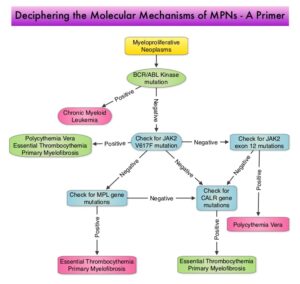
Overactivation of the JAK-STAT signal transduction pathway caused by V617F mutation has been implicated in majority of the patients with P. Vera. This pathway normally is responsible for passing information from outside the cell through the cell membrane to the DNA in the nucleus for gene transcription. Janus Kinase (JAK) family of tyrosine kinases are cytoplasmic proteins and include JAK1, JAK2, JAK3 and TYK2. JAK1 helps propagate the signaling of inflammatory cytokines whereas JAK2 is essential for growth and differentiation of hematopoietic stem cells. These tyrosine kinases mediate cell signaling by recruiting STATs (Signal Transducer and Activator of Transcription), with resulting modulation of gene expression. In patients with P. Vera, the aberrant myeloproliferation is the result of dysregulated JAK2-STAT signaling as well as excess production of inflammatory cytokines, associated with this abnormal signaling. JAK2 mutations such as JAK2 V617F are seen in approximately 95% of patients with P. Vera.Molecular-Mechanisms-of-MPNs

Studies have shown that JAK2 mutations that result in the overproduction of erythrocytes, leukocytes, and platelets in P. Vera also promote direct activation of leukocytes and platelets. Activated platelets and leukocytes bind to each other and activate endothelial cells, which may in turn contribute to the prothrombotic state. The prospective CYTO-PV trial published in 2011, established that maintaining hematocrit less than 45% through phlebotomies and/or cytoreductive drugs significantly decreased the risk of thrombotic events in P. Vera patients. Even though several retrospective analyses strongly suggest an association between leukocytosis and thrombosis and leukocytosis particularly at the time of the thrombotic event in P. Vera patients, no prospective trial has been conducted to assess the impact of WBC counts on thrombotic risk in P. Vera.

The REVEAL study is a large, real-world, multicenter, prospective, noninterventional, observational study, in which patients with P. Vera from US community practice and academic centers were enrolled, to evaluate the association between elevated blood counts and occurrence of thrombotic events in patients with P. Vera, using data from the REVEAL study.

This study analyzed the data of 2271 eligible patients for this analysis (78% high risk and 22% low risk). The median patient age was 66 years and 54% were male. The median disease duration was 4.1 years, 20% had a history of thrombotic events and majority of patients (53%) were receiving Hydroxyurea. Patient data was collected at diagnosis, at a 6-month period, and during follow up, 3 years from last patient enrollment, between July 2014 and August 2019 and the researchers analyzed the association between blood counts and thrombotic events. Out of 106 patients who had thrombotic events, 30 had arterial thrombotic events, most commonly, Transient Ischemic Attack and 76 had venous thrombotic events, most commonly, Deep Vein Thrombosis.

It was noted that hematocrit greater than 45% versus 45% or less (P=0.0028), WBC more than 11×109/L versus 11×109/L or less (P<0.0001), and Platelet counts more than 400×109/L versus 400×109/L or less (P=0.0170) were each associated with increased risk of thrombotic events. A WBC count of 11×109/L or more was associated with the highest thrombotic event risk compared with WBC count less than 7×10⁹/L (P<0.0001).  When the HCT was controlled at 45% or less, an elevated WBC count (more than 12 × 109/L) was significantly associated with increased risk of thromboembolism (HR=1.95; P=0.03). In all models analyzed, advanced age and history of thrombotic events, were associated with increased thrombotic event risk.

The authors concluded that in this analysis of the largest real-world cohort of P. Vera patients to date, hematocrit more than 45%, as well as WBC more than 11×109/L and Platelet counts more than 400×109/L, were each associated with increased risk of thrombotic events. WBC more than 12×109/L was significantly associated with increased thrombotic risk, even when the HCT was controlled, suggesting that thrombotic risk may be reduced by controlling both the WBC count and HCT level. The authors added that these data support the incorporation of blood count values into risk stratification and treatment strategies for patients with P. Vera in clinical practice, and to move beyond the conventional risk model.

A Real-World Evaluation of the Association between Elevated Blood Counts and Thrombotic Events in Polycythemia Vera: An Analysis of Data from the REVEAL Study). Gerds AT, Mesa RA, Burke JM, et al. Presented at: 2022 European Hematology Association Congress; June 10, 2022; Vienna, AT. Poster # P1062.

Association Between Polycystic Ovary Syndrome and Risk of Pancreatic Cancer

SUMMARY: The American Cancer Society estimates that in 2022, about 62,210 people will be diagnosed with pancreatic cancer and 49,830 people will die of the disease. Pancreatic cancer is the fourth most common cause of cancer-related deaths in the United States and Western Europe. Unfortunately, unlike other malignancies, very little progress has been made and outcome for patients with advanced pancreatic cancer has been dismal, with a 5-year survival rate for metastatic pancreatic cancer of approximately 10%. Pancreatic cancer has surpassed breast cancer as the third leading cause of cancer death in the United States and is on track to surpass colorectal cancer, to move to the second leading cause of cancer related deaths in the United States.

Majority of patients with pancreatic cancer (80% of cases) are diagnosed at an advanced stage, and are not amenable to curative surgical resection, at the time of diagnosis. The current treatment regimens for advanced disease have proved ineffective, conferring a median Overall Survival (OS) of 6-8 months. Clinical surveillance is recommended for individuals at high risk, although pancreatic cancer has relatively few established risk factors.

Polycystic Ovary Syndrome (PCOS) is a hormone disorder affecting 7-10% of women of childbearing age and is the most common cause of infertility. In the United States, an estimated 5-6 million women have PCOS and is the most common hormonal disorder among women of reproductive age. The etiology of PCOS is unknown and the signs and symptoms of PCOS have been attributed to abnormal levels of Luteinizing Hormone (LH) and androgen excess, which interfere with normal function of the ovaries. Hyperinsulinemia and insulin resistance can lead to the development of Type 2 diabetes and worsen androgen excess. A previously published study using Swedish register data suggested several carcinogenic processes associated with PCOS, with a 3.4-fold increased risk of pancreatic cancer, in small group of women with PCOS (JAMA Oncol. 2019;5(1):106-107).

To confirm these findings, the authors conducted a case-control study using data from the Memorial Sloan Kettering Cancer Center Pancreatic Tumor Registry. The study population included 446 women (N=446) who were 21 years of age or older and had pathologically or cytologically confirmed pancreatic adenocarcinoma. The control group consisted of visitors accompanying patients to Memorial Sloan Kettering Cancer Center clinics or spouses of patients with pancreatic cancer with no personal history of cancer (N=209). The researchers followed STROBE (Strengthening the Reporting of Observational Studies in Epidemiology) guideline to report their observational research findings and used multivariable logistic regression to estimate Odds Ratios (ORs) and 95% Confidence Intervals (CIs) for the association between PCOS and risk of pancreatic cancer. The median age of the study population was 60 years.

The researchers noted a positive association between PCOS and risk of pancreatic cancer, after adjusting for age, race and ethnicity, BMI, and estrogen use (OR=1.88 – 88% more likely). The Odds Ratio remained significant at 1.78 after adjusting for potential mediator Type 2 diabetes, in addition to all the above factors. Excluding those with pancreatic cancer diagnosed less than 5 years after PCOS diagnosis, history of pancreatitis and a history of familial pancreatic cancer did not materially alter risk estimates. These findings suggested that this association of PCOS and risk of pancreatic cancer was independent of BMI, and not driven by Type 2 diabetes or other variables. The researchers hypothesized that these data suggest some individuals may have unknown metabolic derangements that may underly the development of both conditions.

The authors concluded that in this case-control study, PCOS was strongly associated with increased risk of pancreatic cancer. They added that diagnosis of PCOS may warrant increased education and clinical vigilance for pancreatic cancer, although prospective studies are needed to examine underlying biologic mechanisms and confirm this study findings.

Association Between Polycystic Ovary Syndrome and Risk of Pancreatic Cancer. Peeri NC, Landicino MV, Saldia CA, et al. JAMA Oncol. Published online October 6, 2022. doi:10.1001/jamaoncol.2022.4540

Lung Cancer Screening with Low Dose CT Associated with Favorable Stage Shift and Improved Survival

SUMMARY: The American Cancer Society estimates that for 2022, about 236,740 new cases of lung cancer will be diagnosed and 135,360 patients will die of the disease. Lung cancer is the leading cause of cancer-related mortality in the United States. Non-Small Cell Lung Cancer (NSCLC) accounts for approximately 85% of all lung cancers and Adenocarcinoma now is the most frequent histologic subtype of lung cancer.

In the National Lung Screening Trial (NLST) with Low Dose CT (LDCT) screening for lung cancer, there was a 20% reduction in mortality. Following the publication of the results of NLST, and NCCN issued guideline in 2011, the United States Preventive Services Task Force (USPSTF) recommended Lung Cancer screening with Low Dose CT scan in high risk patients. The CMS in 2015 determined that there was sufficient evidence to reimburse for this preventive service. The USPSTF expanded the criteria for Lung Cancer screening in 2021 and recommended annual screening with Low-Dose CT for adults aged 50 to 80 years who have a 20 pack-year smoking history and currently smoke or have quit within the past 15 years. The new USPSTF 2021 criteria were given a B recommendation, as there was additional research needed, to improve uptake of LDCT screening and to develop biomarkers to more accurately identify individuals, who would benefit from screening.

Approximately 15% of patients present with early stage (T1-2 N0) disease, and these numbers are likely to increase with the implementation of Lung Cancer screening programs. Surgical resection is the primary treatment for approximately 30% of patients with NSCLC who present with early Stage (I–IIIA) disease. In spite of the favorable stage shift as a result of lung cancer screening, low Health Care Provider knowledge of the lung cancer screening guidelines represents a potential barrier to implementation, and no clinical trials have shown these favorable benefits in a real world setting.

The authors in this study evaluated whether the introduction of Low Dose CT screening in 2013 resulted in an increase in the percentage of Stage I NSCLC diagnosed among patients potentially eligible for screening, along with an increase in median all cause survival among these patients, and whether any effects on stage extend to the entire study population or only select population groups. The researchers analyzed data from two large comprehensive US cancer registries-the National Cancer Database and the Surveillance Epidemiology End Results (SEER) program database using a quasi-experimental observational design. A total of 763 474 patients were identified for analysis in this study. They included those who were diagnosed as having NSCLC between 2010 and 2018 and who would have been eligible for screening by age criteria (age 55-79 years) and a comparator NSCLC patient cohort who would have been ineligible for screening (age 45-55). The authors then compared the rate of change in the percentage of patients with Stage I cancer at diagnosis between 2010 and 2018.

It was noted that among the screen eligible cohort of NSCLC patients, the percentage of patients with Stage I disease at diagnosis increased by 3.9% each year from 2014, following a minor change from 2010 to 2013. The rate of increase in Stage I diagnoses was more rapid in high lung cancer screening states. These findings however were not seen in the younger, screening ineligible patients. These results consistently noted across multiple analyses.

The median all cause survival of screening eligible patients aged 55-80 years increased at 11.9% per year from 2014 to 2018 (from 19.7 to 28.2 months). In multivariable adjusted analysis, the hazard of death decreased significantly faster after 2014 compared with before 2014 (P<0.001).

Disparities were however noted, and the benefits from this significant shift in the stage of the disease was not realized in racial or ethnic minority groups and those living in lower income or less educated regions. By 2018, Stage I NSCLC was the predominant diagnosis among non-Hispanic white people, whereas the economically deprived group of patients, were more likely to have Stage IV disease at diagnosis. Increases in the detection of early stage lung cancer in the US from 2014 to 2018 led to an estimated 10,100 averted deaths.

It was concluded from this study that although the adoption of lung cancer screening has been slow nationwide, this study indicated the beneficial effect of lung cancer screening and a recent stage shift toward Stage I NSCLC, with improved survival, following the introduction of lung cancer screening. This study also highlighted the disparities in the stage of lung cancer diagnosed between patient populations, reinforcing the need for equitable access to screening in the US.

Association of computed tomography screening with lung cancer stage shift and survival in the United States: quasi-experimental study. Potter AL, Rosenstein AL, Kiang MV, et al. BMJ 2022; 376 doi: https://doi.org/10.1136/bmj-2021-069008 (Published 30 March 2022)

OPDIVO® (nivolumab) for the Adjuvant Treatment of High-Risk Urothelial Carcinoma*

*Urothelial carcinoma at high risk of recurrence after undergoing radical resection.
Written by: Terence Friedlander, MD
Professor of Medicine, Division of Hematology/Oncology, Zuckerberg San Francisco General Hospital, Helen Diller Family Comprehensive Cancer Center, University of California, San Francisco

Content sponsored by: Bristol Myers Squibb
Dr Friedlander is a paid consultant for BMS and was compensated for his contribution in drafting this content.

Overview of High-Risk Urothelial Carcinoma*
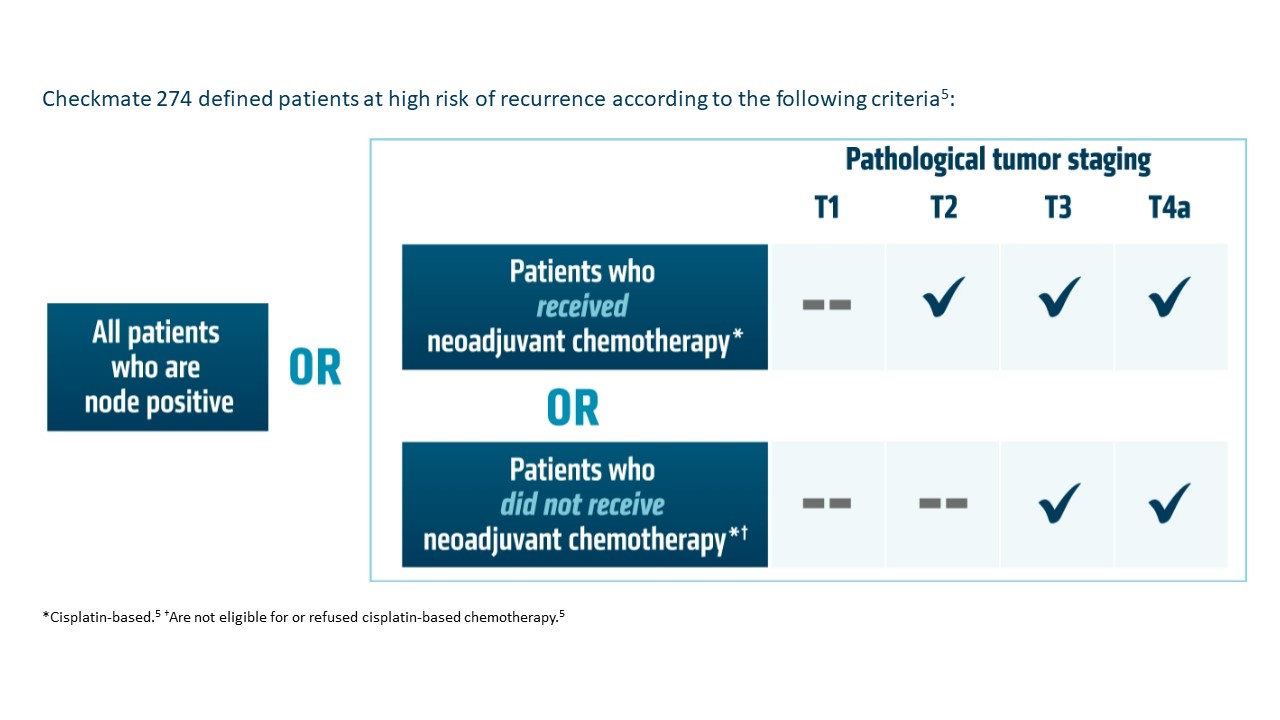
Currently, radical resection with or without perioperative therapy is the standard of care for treating high-risk urothelial carcinoma (UC).1* However, there is still a high chance of recurrence within 2 years of radical resection, with less favorable survival rates for the high-risk patient population.1 While neoadjuvant therapy has an established role in treating high-risk UC,* data are less clear regarding the role of adjuvant therapy.2 In a retrospective observational cohort study of patients 65 years or older with UC at high risk of recurrence after radical resection, including patients who received neoadjuvant chemotherapy, median disease-free survival (mDFS) was determined to be 13.5 months.1 Cisplatin-based chemotherapy is the neoadjuvant standard of care, but prior to 2021 there were no FDA-approved adjuvant therapy options.1-3 Studies have shown that adjuvant chemotherapy may delay recurrence and improve overall survival (OS), but these studies have not definitively shown a survival benefit, largely due to inadequate sample sizes.2 Additionally, approximately 50% of patients are ineligible for cisplatin-based treatment.1 As a result, there is a high unmet need for this difficult-to-treat population, and it is important for the urologist, oncologist, and patient to discuss and align on perioperative treatments at the time of diagnosis and early in the patient journey.1,2,4 Entering the adjuvant treatment landscape, immune checkpoint inhibitors may be an additional treatment option for HCPs to consider for their patients with high-risk UC.1,2*

Adjuvant OPDIVO in High-Risk Urothelial Carcinoma*
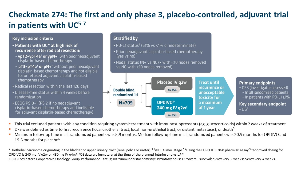
OPDIVO is approved and indicated for the adjuvant treatment of adult patients with UC who are at high risk of recurrence after undergoing radical resection, regardless of prior neoadjuvant chemotherapy, nodal involvement, or PD-L1 status.5 The approval is based on Checkmate 274, a phase 3, multicenter, double-blind, randomized trial of adjuvant OPDIVO versus placebo.6 More information on the study design can be found in the images below. Baseline characteristics were balanced across treatment arms.6
Checkmate 274Important Safety Information
Select Important Safety Information
In Checkmate 274, serious adverse reactions occurred in 30% of OPDIVO patients. The most frequent serious adverse reaction reported in ≥2% of patients was urinary tract infection. Fatal adverse reactions occurred in 1% of patients; these included events of pneumonitis (0.6%). The most-common adverse reactions reported in ≥20% of patients were rash, fatigue, diarrhea, pruritus, musculoskeletal pain, and UTI. OPDIVO was discontinued or delayed due to adverse reactions in 18% and 33% of patients, respectively.5

OPDIVO is associated with the following Warnings and Precautions: severe and fatal immune-mediated adverse reactions including pneumonitis, colitis, hepatitis and hepatotoxicity, endocrinopathies, nephritis with renal dysfunction, dermatologic adverse reactions, other immune-mediated adverse reactions; infusion-related reactions; complications of allogeneic hematopoietic stem cell transplantation; embryo-fetal toxicity; and increased mortality in patients with multiple myeloma when OPDIVO is added to a thalidomide analogue and dexamethasone, which is not recommended outside of controlled clinical trials.

OPDIVO may cause severe infusion-related reactions. In patients who received OPDIVO as a 60-minute intravenous infusion, infusion-related reactions occurred in 6.4% (127/1994) of patients.5 For additional information regarding infusion-related reactions, please see Important Safety Information for OPDIVO.
Checkmate-274-Defined-High-Risk-PatientsDouble-median-DFS-with-OPDIVO
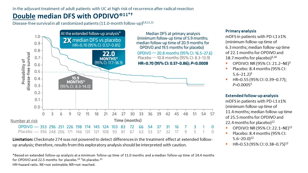
Checkmate 274 was not powered to detect differences in the treatment effect at extended follow-up analysis; therefore, results from this exploratory analysis should be interpreted with caution.

Adjuvant OPDIVO demonstrated superior disease-free survival (DFS) compared with placebo at the primary analysis (minimum follow-up of 5.9 months).5,6 Median DFS was 20.8 months with OPDIVO versus 10.8 months with placebo (HR=0.70 [95% CI: 0.57–0.86];P=0.0008).5 OS was also evaluated as a secondary endpoint, but at the time of the planned interim analysis, these data were immature with 33% of deaths in the ITT population; in the UTUC subpopulation, 37 deaths occurred, 20 of which occurred with OPDIVO versus 17 with placebo.5 Although the subgroup analyses were not statistically powered, for patients with prior neoadjuvant cisplatin therapy (n=308), the DFS hazard ratio was 0.52 [95% CI: 0.38–0.71] and for patients without prior neoadjuvant cisplatin therapy (n=401), the DFS hazard ratio was 0.92 [95% CI: 0.69–1.21].6 In additional exploratory subgroup analyses, no improvement in DFS was observed with nivolumab compared to placebo in patients with UTUC (n=149) the unstratified DFS hazard ratio was 1.15 (95% CI: 0.74–1.80); in patients with PD-L1 expression of <1% (n=414), the unstratified DFS hazard ratio was 0.83 (95% CI: 0.64–1.08).5

At the extended follow-up analysis (minimum follow-up of 11.0 months), mDFS was doubled with adjuvant OPDIVO compared with placebo. Median DFS was 22.0 months with OPDIVO versus 10.9 months with placebo (HR=0.70 [95% CI: 0.57–0.85]).12

Summary/conclusions
Given the high unmet need in this difficult-to-treat population, the call for approved adjuvant treatment options continues to rise.1,2 Adjuvant OPDIVO offers a chance to change the future for patients with high-risk UC as the only FDA-approved adjuvant option for adult patients with UC at high risk of recurrence after radical resection regardless of prior neoadjuvant chemotherapy, nodal involvement, or PD-L1 status.5,6,12 In Checkmate 274, OPDIVO significantly extended mDFS at the time of primary analysis and doubled mDFS at the time of extended follow-up analysis.5,6,12 Further data will be generated for the secondary endpoint of OS, which may provide greater insight into the efficacy of OPDIVO in this context.6,8 Given the clinical profile of Checkmate 274 and subsequent FDA approval, OPDIVO may help extend DFS for appropriate patients in need of treatment in the adjuvant UC setting.5,6,12

*Urothelial carcinoma at high risk of recurrence after undergoing radical resection.

Additional Definitions
CI=confidence interval; HCP=healthcare provider; HR=hazard ratio; ITT=intent to treat; PD-L1=programmed death ligand 1; UTUC=upper tract urothelial carcinoma.

Indication
OPDIVO® (nivolumab), as a single agent, is indicated for the adjuvant treatment of adult patients with urothelial carcinoma (UC) who are at high risk of recurrence after undergoing radical resection of UC.

Important Safety Information
Severe and Fatal Immune-Mediated Adverse Reactions
Immune-mediated adverse reactions listed herein may not include all possible severe and fatal immune-mediated adverse reactions.

Immune-mediated adverse reactions, which may be severe or fatal, can occur in any organ system or tissue. While immune-mediated adverse reactions usually manifest during treatment, they can also occur after discontinuation of OPDIVO. Early identification and management are essential to ensure safe use of OPDIVO. Monitor for signs and symptoms that may be clinical manifestations of underlying immune-mediated adverse reactions. Evaluate clinical chemistries including liver enzymes, creatinine, and thyroid function at baseline and periodically during treatment with OPDIVO. In cases of suspected immune-mediated adverse reactions, initiate appropriate workup to exclude alternative etiologies, including infection. Institute medical management promptly, including specialty consultation as appropriate.

Withhold or permanently discontinue OPDIVO depending on severity (please see section 2 Dosage and Administration in the accompanying Full Prescribing Information). In general, if OPDIVO interruption or discontinuation is required, administer systemic corticosteroid therapy (1 to 2 mg/kg/day prednisone or equivalent) until improvement to Grade 1 or less. Upon improvement to Grade 1 or less, initiate corticosteroid taper and continue to taper over at least 1 month. Consider administration of other systemic immunosuppressants in patients whose immune-mediated adverse reactions are not controlled with corticosteroid therapy. Toxicity management guidelines for adverse reactions that do not necessarily require systemic steroids (e.g., endocrinopathies and dermatologic reactions) are discussed below.

Immune-Mediated Pneumonitis
OPDIVO can cause immune-mediated pneumonitis. The incidence of pneumonitis is higher in patients who have received prior thoracic radiation. In patients receiving OPDIVO monotherapy, immune-mediated pneumonitis occurred in 3.1% (61/1994) of patients, including Grade 4 (<0.1%), Grade 3 (0.9%), and Grade 2 (2.1%).

Immune-Mediated Colitis
OPDIVO can cause immune-mediated colitis. A common symptom included in the definition of colitis was diarrhea. Cytomegalovirus (CMV) infection/reactivation has been reported in patients with corticosteroid-refractory immune-mediated colitis. In cases of corticosteroid-refractory colitis, consider repeating infectious workup to exclude alternative etiologies. In patients receiving OPDIVO monotherapy, immune-mediated colitis occurred in 2.9% (58/1994) of patients, including Grade 3 (1.7%) and Grade 2 (1%).

Immune-Mediated Hepatitis and Hepatotoxicity
OPDIVO can cause immune-mediated hepatitis. In patients receiving OPDIVO monotherapy, immune-mediated hepatitis occurred in 1.8% (35/1994) of patients, including Grade 4 (0.2%), Grade 3 (1.3%), and Grade 2 (0.4%).

Immune-Mediated Endocrinopathies
OPDIVO can cause primary or secondary adrenal insufficiency, immune-mediated hypophysitis, immune- mediated thyroid disorders, and Type 1 diabetes mellitus, which can present with diabetic ketoacidosis. Withhold OPDIVO depending on severity (please see section 2 Dosage and Administration in the accompanying Full Prescribing Information). For Grade 2 or higher adrenal insufficiency, initiate symptomatic treatment, including hormone replacement as clinically indicated. Hypophysitis can present with acute symptoms associated with mass effect such as headache, photophobia, or visual field defects. Hypophysitis can cause hypopituitarism; initiate hormone replacement as clinically indicated. Thyroiditis can present with or without endocrinopathy. Hypothyroidism can follow hyperthyroidism; initiate hormone replacement or medical management as clinically indicated. Monitor patients for hyperglycemia or other signs and symptoms of diabetes; initiate treatment with insulin as clinically indicated.

In patients receiving OPDIVO monotherapy, adrenal insufficiency occurred in 1% (20/1994), including Grade 3 (0.4%) and Grade 2 (0.6%).

In patients receiving OPDIVO monotherapy, hypophysitis occurred in 0.6% (12/1994) of patients, including Grade 3 (0.2%) and Grade 2 (0.3%).

In patients receiving OPDIVO monotherapy, thyroiditis occurred in 0.6% (12/1994) of patients, including Grade 2 (0.2%).

In patients receiving OPDIVO monotherapy, hyperthyroidism occurred in 2.7% (54/1994) of patients, including Grade 3 (<0.1%) and Grade 2 (1.2%).

In patients receiving OPDIVO monotherapy, hypothyroidism occurred in 8% (163/1994) of patients, including Grade 3 (0.2%) and Grade 2 (4.8%).

In patients receiving OPDIVO monotherapy, diabetes occurred in 0.9% (17/1994) of patients, including Grade 3 (0.4%) and Grade 2 (0.3%), and 2 cases of diabetic ketoacidosis.

Immune-Mediated Nephritis with Renal Dysfunction
OPDIVO can cause immune-mediated nephritis. In patients receiving OPDIVO monotherapy, immune-mediated nephritis and renal dysfunction occurred in 1.2% (23/1994) of patients, including Grade 4 (<0.1%), Grade 3 (0.5%), and Grade 2 (0.6%).

Immune-Mediated Dermatologic Adverse Reactions
OPDIVO can cause immune-mediated rash or dermatitis. Exfoliative dermatitis, including Stevens-Johnson syndrome (SJS), toxic epidermal necrolysis (TEN), and drug rash with eosinophilia and systemic symptoms (DRESS) has occurred with PD-1/PD-L1 blocking antibodies. Topical emollients and/or topical corticosteroids may be adequate to treat mild to moderate nonexfoliative rashes.

Withhold or permanently discontinue OPDIVO depending on severity (please see section 2 Dosage and Administration in the accompanying Full Prescribing Information).

In patients receiving OPDIVO monotherapy, immune-mediated rash occurred in 9% (171/1994) of patients, including Grade 3 (1.1%) and Grade 2 (2.2%).

Other Immune-Mediated Adverse Reactions
The following clinically significant immune-mediated adverse reactions occurred at an incidence of <1% (unless otherwise noted) in patients who received OPDIVO monotherapy or were reported with the use of other PD-1/PD- L1 blocking antibodies. Severe or fatal cases have been reported for some of these adverse reactions: cardiac/vascular: myocarditis, pericarditis, vasculitis; nervous system: meningitis, encephalitis, myelitis and demyelination, myasthenic syndrome/myasthenia gravis (including exacerbation), Guillain-Barré syndrome, nerve paresis, autoimmune neuropathy; ocular: uveitis, iritis, and other ocular inflammatory toxicities can occur; gastrointestinal: pancreatitis to include increases in serum amylase and lipase levels, gastritis, duodenitis; musculoskeletal and connective tissue: myositis/polymyositis, rhabdomyolysis, and associated sequelae including renal failure, arthritis, polymyalgia rheumatica; endocrine: hypoparathyroidism; other (hematologic/immune): hemolytic anemia, aplastic anemia, hemophagocytic lymphohistiocytosis (HLH), systemic inflammatory response syndrome, histiocytic necrotizing lymphadenitis (Kikuchi lymphadenitis), sarcoidosis, immune thrombocytopenic purpura, solid organ transplant rejection.

Some ocular IMAR cases can be associated with retinal detachment. Various grades of visual impairment, including blindness, can occur. If uveitis occurs in combination with other immune-mediated adverse reactions, consider a Vogt-Koyanagi-Harada–like syndrome, which has been observed in patients receiving OPDIVO, as this may require treatment with systemic corticosteroids to reduce the risk of permanent vision loss.

Infusion-Related Reactions
OPDIVO can cause severe infusion-related reactions. Discontinue OPDIVO in patients with severe (Grade 3) or life-threatening (Grade 4) infusion-related reactions. Interrupt or slow the rate of infusion in patients with mild (Grade 1) or moderate (Grade 2) infusion-related reactions. In patients receiving OPDIVO monotherapy as a 60- minute infusion, infusion-related reactions occurred in 6.4% (127/1994) of patients. In a separate trial in which patients received OPDIVO monotherapy as a 60-minute infusion or a 30-minute infusion, infusion-related reactions occurred in 2.2% (8/368) and 2.7% (10/369) of patients, respectively. Additionally, 0.5% (2/368) and 1.4% (5/369) of patients, respectively, experienced adverse reactions within 48 hours of infusion that led to dose delay, permanent discontinuation or withholding of OPDIVO.

Complications of Allogeneic Hematopoietic Stem Cell Transplantation
Fatal and other serious complications can occur in patients who receive allogeneic hematopoietic stem cell transplantation (HSCT) before or after being treated with OPDIVO. Transplant-related complications include hyperacute graft-versus-host-disease (GVHD), acute GVHD, chronic GVHD, hepatic veno-occlusive disease (VOD) after reduced intensity conditioning, and steroid-requiring febrile syndrome (without an identified infectious cause). These complications may occur despite intervening therapy between OPDIVO and allogeneic HSCT.

Follow patients closely for evidence of transplant-related complications and intervene promptly. Consider the benefit versus risks of treatment with OPDIVO prior to or after an allogeneic HSCT.

Embryo-Fetal Toxicity
Based on its mechanism of action and findings from animal studies, OPDIVO can cause fetal harm when administered to a pregnant woman. Advise pregnant women of the potential risk to a fetus. Advise females of reproductive potential to use effective contraception during treatment with OPDIVO and for at least 5 months after the last dose.

Increased Mortality in Patients with Multiple Myeloma when OPDIVO is Added to a Thalidomide Analogue and Dexamethasone
In randomized clinical trials in patients with multiple myeloma, the addition of OPDIVO to a thalidomide analogue plus dexamethasone resulted in increased mortality. Treatment of patients with multiple myeloma with a PD-1 or PD-L1 blocking antibody in combination with a thalidomide analogue plus dexamethasone is not recommended outside of controlled clinical trials.

Lactation
There are no data on the presence of OPDIVO in human milk, the effects on the breastfed child, or the effects on milk production. Because of the potential for serious adverse reactions in breastfed children, advise women not to breastfeed during treatment and for 5 months after the last dose.

Serious Adverse Reactions
In Checkmate 274, serious adverse reactions occurred in 30% of patients receiving OPDIVO (n=351). The most frequent serious adverse reaction reported in ≥2% of patients receiving OPDIVO was urinary tract infection. Fatal adverse reactions occurred in 1% of patients; these included events of pneumonitis (0.6%).

Common Adverse Reactions
In Checkmate 274, the most common adverse reactions (≥20%) reported in patients receiving OPDIVO (n=351) were rash (36%), fatigue (36%), diarrhea (30%), pruritus (30%), musculoskeletal pain (28%), and urinary tract infection (22%).

Please see US Full Prescribing Information for OPDIVO.

References
1. Drakaki A, Pantuck A, Mhatre SK, et al. “Real-world” outcomes and prognostic indicators among patients with high-risk muscle-invasive urothelial carcinoma. Urol Oncol. 2021;39:76.e15-76.e22.
2. Referenced without permission from the NCCN Clinical Practice Guidelines in Oncology (NCCN Guidelines®) for Bladder Cancer V.2.2022. © National Comprehensive Cancer Network, Inc. 2022. All rights reserved. Accessed August 4, 2022. To view the most recent and complete version of the guidelines, go online to NCCN.org. NCCN makes no warranties of any kind whatsoever regarding their content, use or application and disclaims any responsibility for their application or use in any way.
3. Apolo AB, Msaouel P, Niglio S, et al. Evolving Role of Adjuvant Systemic Therapy for Kidney and Urothelial Cancers. Am Soc Clin Oncol Educ Book. 2022;42:1-16. doi:10.1200/EDBK_350829.
4. Nayan M, Bhindi B, Yu JL, et al. The initiation of a multidisciplinary bladder cancer clinic and the uptake of neoadjuvant chemotherapy: A time-series analysis. Can Urol Assoc J. 2016;10(1-2):25-30.
5. OPDIVO [package insert]. Princeton, NJ: Bristol-Myers Squibb Company.
6. Bajorin DF, Witjes JA, Gschwend JE, et al. Adjuvant nivolumab versus placebo in muscle-invasive urothelial carcinoma. N Engl J Med. 2021;384(22):2102-2114.
7. Bajorin DF, Witjes JA, Gschwend JE, et al. First results from the phase 3 CheckMate 274 trial of adjuvant nivolumab versus placebo in patients who underwent radical surgery for high-risk muscle-invasive urothelial carcinoma. Oral presentation at ASCO GU 2021. Abstract 391.
8. Bajorin DF, Witjes JA, Gschwend JE, et al. Adjuvant nivolumab versus placebo in muscle-invasive urothelial carcinoma. N Engl J Med. 2021;384(22):2102-2114 [supplementary appendix].
9. American Cancer Society. Bladder cancer early detection, diagnosis, and staging. Accessed August 5, 2022. https://www.cancer.org/content/dam/CRC/PDF/Public/8559.00.pdf.
10. Data on file. NIVO 639. Princeton, NJ: Bristol-Myers Squibb Company; 2021.
11. Witjes JA, Bajorin DF, Galsky MD, et al. Results for patients with muscle-invasive bladder cancer in the CheckMate 274 trial. Poster presentation at ASCO 2022. Abstract 4585.
12. Galsky MD, Witjes JA, Gschwend JE, et al. Disease-free survival with longer follow-up from the phase 3 CheckMate 274 trial of adjuvant nivolumab in patients who underwent surgery for high-risk muscle-invasive urothelial carcinoma. Oral presentation at the American Urological Association (AUA) Annual Meeting 2022. Abstract 22-3807.

© 2022 Bristol-Myers Squibb Company. OPDIVO® and the related logos are trademarks of Bristol-Myers Squibb Company. 1506-US-2200368 8/22

FDA Grants Accelerated Approval to LYTGOBI® for Cholangiocarcinoma

SUMMARY: The FDA on September 30, 2022, granted accelerated approval to LYTGOBI® (Futibatinib) for adult patients with previously treated, unresectable, locally advanced, or metastatic intrahepatic cholangiocarcinoma harboring Fibroblast Growth Factor Receptor 2 (FGFR2) gene fusions or other rearrangements. Bile Tract cancer (Cholangiocarcinoma) is a rare, heterogenous cancer, and comprises about 30% of all primary liver tumors and includes both intrahepatic and extrahepatic bile duct cancers. Klatskin tumor is a type of Cholangiocarcinoma that begins in the hilum, at the junction of the left and right bile ducts. It is the most common type of Cholangiocarcinoma, accounting for more than half of all cases. About 8,000 people in the US are diagnosed with Cholangiocarcinoma each year and approximately 20% of the cases are suitable for surgical resection. The 5-year survival among those with advanced stage disease is less than 10%, with limited progress made over the past two decades.

Approximately 75% of patients are diagnosed with late-stage disease, and are often treated with Gemcitabine plus Cisplatin, based on the findings of the ABC-02 study. Second line treatment options include FOLFOX regimen, which is associated with a Response Rate of about 5%, median Progression Free Survival (PFS) of about 4 months, and median Overall Survival (OS) of about 6 months. There is therefore an unmet need for new effective therapies.

FGFRs (Fibroblast Growth Factor Receptors) play an important role in tumor cell proliferation and survival, migration, and angiogenesis. Activating fusions, rearrangements, translocations, and gene amplifications in FGFRs result in dysregulation of FGFR signaling, and may contribute to the pathogenesis of various cancers, including Cholangiocarcinoma. FGFR2 fusions or rearrangements occur almost exclusively in intrahepatic Cholangiocarcinoma, where they are observed in 10-20% of patients, and have been identified as oncogenic drivers. Futibatinib is a highly selective, irreversible FGFR1-4 inhibitor, and demonstrated tolerability and preliminary evidence of clinical efficacy in patients with intrahepatic cholangiocarcinoma.

The present FDA approval was based on the results from the pivotal FOENIX-CCA2 trial (NCT02052778), which is a global, multicenter, open-label, single-arm study that enrolled 103 patients with previously treated, unresectable, locally advanced or metastatic intrahepatic cholangiocarcinoma, harboring a FGFR2 gene fusion or other rearrangement. The presence of FGFR2 fusions or other rearrangements was determined using Next Generation Sequencing testing. Patients received Futibatinib 20 mg orally once daily until disease progression or unacceptable toxicity. The median age was 58 years, 53% had an ECOG Performance Status of 1, all patients had prior anticancer therapy, with 27% receiving prior radiotherapy. FGFR2 fusions were observed in 78% of patients and 22% had a rearrangement. The median time from prior anticancer therapy to the first Futibatinib dose was 1.5 months. The Primary endpoint was Objective Response Rate (ORR) by Independent Central Review. Secondary endpoints were Duration of Response (DOR), Disease Control Rate (DCR), Progression Free Survival (PFS), Overall Survival (OS), Safety, and Patient-Reported Outcomes. At the primary analysis of this trial, an Objective Response Rate of 41.7% was observed, with a median Duration of Response of 9.7 months. The researchers herein reported updated efficacy, including mature Overall Survival, and safety data from the final analysis, with an additional 8 months of follow up.

At a median follow up of 25 months, the median number of treatment cycles was 13.0 and the median treatment duration was 9.1 months. The confirmed Objective Response Rate was 41.7%, like what was noted at the time of primary analysis, and this benefit was consistent across patient subgroups. The Disease Control Rate of 82.5% and was similar as well. The median Duration of Response was 9.5 months, and 74% of responses lasted 6 months or more. The median PFS was 8.9 months, with a 12-month PFS rate of 35%. The median Overall Survival was 20 months, with a 12-month Overall Survival rate of 73%. The most common treatment-related adverse events included hyperphosphatemia (85%), alopecia (33%), dry mouth (30%), diarrhea (28%), dry skin (27%), and fatigue (25%). Approximately 4% of patients discontinued treatment due to adverse events.

The authors concluded that the final analysis of FOENIX-CCA2 study confirmed the results of the primary analysis and reinforced the durable efficacy and continued tolerability of Futibatinib in previously treated patients with advanced/metastatic intrahepatic cholangiocarcinoma harboring FGFR2 fusion/rearrangements. They added that the mature Overall Survival far exceeded historical data in this patient population.

Updated results of the FOENIX-CCA2 trial: Efficacy and safety of futibatinib in intrahepatic cholangiocarcinoma (iCCA) harboring FGFR2 fusions/rearrangements. Goyal L, Meric-Bernstam F, Hollebecque A, et al. J ClinOncol. 2022;40(suppl 16):4009. doi:10.1200/JCO.2022.40.16_suppl.4009

Expansion of Cancer Risk Profile beyond Breast and Ovarian Cancer for BRCA1 and BRCA2 Pathogenic Variants

SUMMARY: DNA damage is a common occurrence in daily life by UV light, ionizing radiation, replication errors, chemical agents, etc. This can result in single and double strand breaks in the DNA structure which must be repaired for cell survival. The vital pathways for DNA repair in a normal cell are BRCA1/BRCA2 and PARP. BRCA1 and BRCA2 genes recognize and repair double strand DNA breaks via Homologous Recombination Repair (HRR) pathway. Homologous Recombination is a type of genetic recombination, and is a DNA repair pathway utilized by cells to accurately repair DNA double-stranded breaks during the S and G2 phases of the cell cycle, and thereby maintain genomic integrity. Homologous Recombination Deficiency (HRD) is noted following mutation of genes involved in HR repair pathway.

BRCA1 and BRCA2 are tumor suppressor genes located on chromosome 17 and chromosome 13 respectively and functional BRCA proteins repair damaged DNA, and play an important role in maintaining cellular genetic integrity. They regulate cell growth and prevent abnormal cell division and development of malignancy. Mutations in these genes predispose an individual to develop malignant tumors.

BRCA mutations can either be inherited (Germline) and present in all individual cells or can be acquired and occur exclusively in the tumor cells (Somatic). Somatic mutations account for a significant portion of overall BRCA1 and BRCA2 aberrations. Loss of BRCA function due to frequent somatic aberrations likely deregulates HR pathway, and other pathways then come in to play, which are less precise and error prone, resulting in the accumulation of additional mutations and chromosomal instability in the cell, with subsequent malignant transformation. Homologous Recombination Deficiency therefore indicates an important loss of DNA repair function.

Pathogenic Variants (PVs) in BRCA1 and BRCA2 (BRCA1/2) are well known to be associated with increased lifetime risk for breast and ovarian cancer in women, and reliable risk estimates are also available and can be as high as 85% and 40% respectively. However, the association of BRCA1 and BRCA2 Pathogenic Variants with cancers other than female breast and ovarian cancers remain uncertain, and these associations have been based on studies with relatively small sample sizes, resulting in imprecise cancer risk estimates. It is therefore important that precise risk estimates are available, in order to optimize clinical management strategies and guidelines for cancer risk management in female and male BRCA1/2 carriers. The NCCN and other guidelines recommend breast and ovarian cancer screening for BRCA1/2 carriers, prostate cancer screening for BRCA2 carriers. Screening is also recommended for pancreatic cancer in BRCA1/2 carriers, but only in the presence of a positive family history of the disease.

The researchers conducted this study to evaluate the association of BRCA1 and BRCA2 pathogenic variants, with additional cancer types and their clinical characteristics associated with pathogenic variant carrier status. For this study, a large-scale registry based sequencing study was performed across 14 common cancer types in 63, 828 patients and 37, 086 controls, whose data were drawn from a Japanese nationwide multi-institutional hospital-based biobank, between 2003 and 2018. In the study group, the median age was 64 years and 42% were female, whereas the median age was 62 years and 47% were female in the control group. Germline pathogenic variants were identified in the BRCA1 and BRCA2 genes by a multiplex Polymerase Chain Reaction-based target sequence method. Associations of (likely) pathogenic variants with each cancer type were assessed by comparing pathogenic variant carrier frequency between patients in each cancer type and controls. Compared with the researchers previous publications for breast, colorectal, pancreatic, and prostate cancers, this study included 14,448 additional controls and 8247 additional cancer cases. These data thus provided a broad view of cancer risks associated with pathogenic variants in BRCA1 and BRCA2 genes.

Pathogenic variants in BRCA1 were significantly associated with increased risk for three other types of cancer types, Biliary tract (Odds Ratio–OR=17.4), Gastric (OR=5.2), and Pancreatic cancer (OR=12.6), in addition to female Breast (OR=16.1) and Ovarian cancer (OR=75.6). Pathogenic variants in BRCA2 increased risk for seven cancer types which included female Breast (OR=10.9), male Breast (OR=67.9), Gastric (OR=4.7), Ovarian (OR=11.3), Pancreatic (OR=10.7), Prostate (OR=4.0), and Esophageal cancer (OR=5.6). Further, Biliary tract, female Breast, Ovarian, and Prostate cancers showed enrichment of carrier patients according to the increased number of reported cancer types in relatives.

The results of this large study suggested that pathogenic variants in BRCA1 and/or BRCA2 are associated with increased risk of biliary tract, gastric, and esophageal cancers, higher than for European populations, granted that these cancers are known to have a higher incidence rate in East Asian countries. Conversely in this study, the cumulative risk of prostate cancer for BRCA2 carriers was lower than that estimated in the UK and Ireland, suggesting that the cumulative risk for each cancer type may be associated with the different incidence rate in each country.

The authors concluded that this study suggested that pathogenic variants in BRCA1 and BRCA2 were associated with the risk of 7 cancer types and is likely broader than that determined from previous analysis of largely European ancestry cohorts. It would therefore be useful to expand indications for genetic testing of individuals with family history of these cancer types.

Expansion of Cancer Risk Profile for BRCA1 and BRCA2 Pathogenic Variants. Momozawa Y, Sasai R, Usui Y, et al. JAMA Oncol. 2022 Apr 14: e220476. doi: 10.1001/jamaoncol.2022.0476 [Epub ahead of print]

FDA Grants Tumor-Agnostic Accelerated Approval to RETEVMO®

SUMMARY: The FDA on September 21, 2022, granted accelerated approval to Selpercatinib (RETEVMO®) for adult patients with locally advanced or metastatic solid tumors with a Rearranged during Transfection (RET) gene fusion that have progressed on or following prior systemic treatment, or who have no satisfactory alternative treatment options. The FDA on the same day also granted Regular approval to Selpercatinib for adult patients with locally advanced or metastatic Non Small Cell Lung Cancer (NSCLC) with a Rearranged during Transfection (RET) gene fusion, as detected by an FDA-approved test. FDA also approved the Oncomine Dx Target (ODxT) Test as a companion diagnostic for Selpercatinib.

In addition to the well characterized gene fusions involving ALK and ROS1 in NSCLC, genetic alterations involving other kinases including EGFR, BRAF, RET, NTRK, are all additional established targetable drivers. These genetic alterations are generally mutually exclusive, with no more than one predominant driver in any given cancer. The hallmark of all these genetic alterations is oncogene addiction, in which cancers are driven primarily, or even exclusively, by aberrant oncogene signaling, and are highly susceptible to small molecule inhibitors.

RET kinase is a transmembrane Receptor Tyrosine Kinase and plays an important role during the development and maintenance of a variety of tissues, including neural and genitourinary tissues. RET signaling activates downstream pathways such as JAK/STAT3 and RAS/RAF/MEK/ERK and leads to cellular proliferation, survival, invasion, and metastasis. Oncogenic alterations to the RET proto-oncogene result in uncontrolled cell growth and enhanced tumor invasiveness. RET alterations include RET rearrangements, leading to RET fusions, and activating point mutations occurring across multiple tumor types. RET fusions have been identified in approximately 2% of NSCLCs, 10-20% of non-medullary thyroid cancers. Activating RET point mutations account for approximately 60% of sporadic Medullary Thyroid Cancers (MTC) and more than 90% of inherited MTCs. Other cancers with documented RET alterations include colorectal, pancreas, breast, and several hematologic malignancies.

Selpercatinib is a highly selective and potent, oral anti-RET Tyrosine Kinase Inhibitor (TKI) designed to inhibit native RET signaling, as well as anticipated acquired resistance mechanisms. Selpercatinib selectively targets wild-type RET as well as various RET mutants and RET-containing fusion products. Additionally, Selpercatinib inhibits Vascular Endothelial Growth Factor Receptor 1 (VEGFR1), VEGFR3, Fibroblast Growth Factor Receptor 1 (FGFR1), FGFR2, and FGFR3. This results in inhibition of cell growth of tumors that exhibit increased RET activity.

The LIBRETTO-001 is the largest open-label, multicenter, Phase I/II trial in patients with advanced solid tumors, including RET fusion-positive solid tumors, RET-mutant Medullary Thyroid Cancers, and other tumors with RET activation, treated with a RET inhibitor. To investigate the efficacy of Selpercatinib, the trial was conducted in 2 parts: Phase 1 (dose escalation) and Phase II (dose expansion). Patients with advanced cancer were eligible, if they have progressed on or were intolerant to available standard therapies, or no standard or available curative therapy existed, or in the opinion of the Investigator, they would be unlikely to tolerate or derive significant clinical benefit from appropriate standard of care therapy, or they declined standard therapy. A dose of 160 mg BID was the recommended Phase II dose. Up to about 850 patients with advanced solid tumors harboring a RET gene alteration in tumor and/or blood were enrolled in 6 different Phase II cohorts, based on tumor type, RET alteration and prior therapy. Identification of RET gene alterations were prospectively determined in local laboratories using either Next Generation Sequencing, Polymerase Chain Reaction, or Fluorescence In Situ Hybridization. The Phase II portion of the trial had a Primary endpoint of Objective Response Rate (ORR) by Blinded Independent Review Committee (BIRC) and Secondary endpoints of Duration of Response, CNS Objective Response Rate, Progression Free Survival (PFS) and safety.

RET Fusion-Positive Solid Tumors

This group included 41 patients and the most common cancers were pancreatic adenocarcinoma (27%), colorectal (24%), salivary (10%), and unknown primary (7%). Majority of the patients (90%) received 2 prior systemic therapies and 32% had received 3 or more. The median age of patients was 50 years, 54% were female, 68% were White, 24% were Asian, and 95% had metastatic disease. RET fusion-positive status was detected in 98% of patients using NGS and 2% using FISH.

The Objective Response Rate was 44%, with 5% Complete Response and 39% Partial Response. The median Duration of response was 24.5 months and 67% of patients had a Duration of Response of 6 months or more.

The NSCLC Cohort

Selpercatinib was previously granted accelerated approval in May 2020 for patients with metastatic RET fusion-positive NSCLC based on initial Overall Response Rate (ORR) and Duration of Response (DOR) among 144 patients enrolled in the LIBRETTO-001 trial. The conversion to regular and traditional FDA approval was based on data from an additional 172 patients and 18 months of additional follow up, to assess durability of response. Patients received Selpercatinib until disease progression or unacceptable toxicity and efficacy was evaluated in a total of 316 patients with locally advanced or metastatic RET fusion-positive NSCLC. The median age of patients was 61 years, 58% were female, 49% were White, 41% were Asian and 97% had metastatic disease. Previously treated patients received a median of two prior systemic therapies and 58% had received prior anti PD 1/PD-L1 therapy.

Among the 69 treatment-naïve patients, the ORR was 84%, with 6% Complete Response and 78% Partial Response. The median Duration of Response was 20.2 months and 50% of patients had a Duration of Response of 12 months or more. Among the 247 previously treated patients, the ORR was 61%, with 7% Complete Response and 54% Partial Response. The median Duration of Response was 28.6 months and 63% of patients had a Duration of Response of 12 months or more.

It is estimated that up to 50% of RET fusion-positive NSCLC patients can have brain metastases, and in the subset of patients with brain metastases (N=21), treatment with Selpercatinib demonstrated a CNS Objective Response Rate of 85%, and 38% of responders had an intracranial Duration of Response of 12 months or greater. The most common toxicities in patients were edema, diarrhea, fatigue, dry mouth, hypertension, abdominal pain, constipation, rash, nausea, and headache.

LIBRETTO-001 is the largest trial ever reported in RET-altered cancer patients and represents an important milestone in the Precision Medicine arena. Selpercatinib is the first and only RET inhibitor to receive both tumor-agnostic accelerated approval and traditional approval in NSCLC, reinforcing its benefits across diverse tumor types.

Selpercatinib in patients with RET fusion–positive non–small-cell lung cancer: updated safety and efficacy from the registrational libretto-001 phase I/II trial.Published September 19, 2022. Drilon A, Subbiah V, Gautschi O, et al. J Clin Oncol. doi:10.1200/JCO.22.00393

https://www.fda.gov/drugs/resources-information-approved-drugs/fda-approves-selpercatinib-locally-advanced-or-metastatic-ret-fusion-positive-solid-tumors

Avoiding Radioiodine Therapy following Thyroidectomy in Patients with Low-Risk Thyroid Cancer

SUMMARY: The American Cancer Society estimates that about 43,800 new cases of thyroid cancer will be diagnosed in the United States in 2022 and about 2,230 patients will die of the disease. Differentiated Thyroid Cancer (DTC) is the most common endocrine malignancy and includes Papillary, Follicular, and Hürthle-cell cancers, with Papillary thyroid cancers accounting for 80% of them. Majority of patients with DTC have clinical Stage I or Stage II disease, with a recurrence rate of less than 5% and cancer-related death rates even lower. Risk factors for recurrence include tumor size, multifocality, capsular or angioinvasion, degree of cervical lymph node involvement, existence of BRAF V600E Mutation, and thyroglobulin levels more than 0.5 ng/mL, after thyroidectomy.

Even though Radioiodine (iodine-131) therapy is not recommended for patients with a unifocal microcarcinoma (10 mm or less in diameter) following thyroidectomy, Radioiodine therapy is generally offered to a majority of patients with low-risk thyroid cancer, both to ablate residual normal thyroid tissue and to treat unresectable persistent disease. The benefits of this intervention however remain controversial. .

The authors conducted a prospective, multicenter, randomized, Phase III Essai Stimulation Ablation 2 (ESTIMABL2) trial involving patients with low-risk thyroid cancer, to assess the non-inferiority of observation versus postoperative Radioiodine therapy, following thyroidectomy. In this study, a total of 776 patients with low-risk Differentiated Thyroid Cancer who were undergoing thyroidectomy were randomly assigned 1:1 to receive ablation with postoperative Radioiodine therapy at a dose of 1.1 GBq (N=389) or no Radioiodine therapy (N=387). Enrolled patients had Differentiated Thyroid Carcinoma (Papillary, Follicular, or Oncocytic/Hürthle-cell cancer), with a multifocal pT1a tumor or a pT1b tumor. None of the patients had regional lymph node involvement, extrathyroidal extension or aggressive histologic subtypes (tall-cell, clear-cell, columnar-cell, and diffuse sclerosing variants of Papillary thyroid cancer, poorly differentiated). The mean patient age was 52 years, and 83% were women, 96% had papillary tumors 81% had pT1b N0 or Nx disease. All patients had normal results on postoperative neck ultrasonography. The follow-up protocol consisted of the measurement of thyroglobulin and thyroglobulin antibodies in all patients at 10 months and yearly thereafter. Ultrasonography of the neck was performed in all patients 10 months and 3 years after randomization. Disease-related events included residual or recurrent disease on neck ultrasonography and a serum thyroglobulin level of more than 1 ng/mL in the group receiving radioiodine and a level of more than 5 ng/mL in the nontreated group. No diagnostic Radioiodine scanning was performed after the whole-body scanning that was performed after therapy. The Primary objective was to assess whether no Radioiodine therapy was noninferior to Radioiodine therapy, with respect to the absence of a composite end point that included functional, structural, and biologic abnormalities, indicating residual or recurrent disease at 3 years.

After 3 years of follow up, there were no clinically meaningful differences in any of the end points between the two groups and the percentage of patients without an event was 95.6% in the no-Radioiodine therapy group and 95.9% in the Radioiodine therapy group, a result that met the noninferiority criteria. Events were more frequent in patients with a postoperative serum thyroglobulin level of more than 1 ng/mL during thyroid hormone treatment. BRAF V600E molecular alterations, which are associated aggressive tumor characteristics, were found in approximately 50% of the samples in each treatment group. The mutational status did not influence event rates in these low-risk patients. No treatment-related adverse events were reported and there was no difference in Quality-of-Life scores between the two groups.

It was concluded that in patients with low-risk thyroid cancer undergoing thyroidectomy, follow up without the use of Radioiodine therapy was noninferior to an ablation strategy with Radioiodine therapy, suggesting that patients with low-risk disease generally do well, regardless of whether they receive Radioiodine therapy.

Thyroidectomy without Radioiodine in Patients with Low-Risk Thyroid Cancer. Leboulleux S, Bournaud C, Chougnet CN, et al. N Engl J Med 2022; 386:923-932