Expansion of Cancer Risk Profile beyond Breast and Ovarian Cancer for BRCA1 and BRCA2 Pathogenic Variants

SUMMARY: DNA damage is a common occurrence in daily life by UV light, ionizing radiation, replication errors, chemical agents, etc. This can result in single and double strand breaks in the DNA structure which must be repaired for cell survival. The vital pathways for DNA repair in a normal cell are BRCA1/BRCA2 and PARP. BRCA1 and BRCA2 genes recognize and repair double strand DNA breaks via Homologous Recombination Repair (HRR) pathway. Homologous Recombination is a type of genetic recombination, and is a DNA repair pathway utilized by cells to accurately repair DNA double-stranded breaks during the S and G2 phases of the cell cycle, and thereby maintain genomic integrity. Homologous Recombination Deficiency (HRD) is noted following mutation of genes involved in HR repair pathway.

BRCA1 and BRCA2 are tumor suppressor genes located on chromosome 17 and chromosome 13 respectively and functional BRCA proteins repair damaged DNA, and play an important role in maintaining cellular genetic integrity. They regulate cell growth and prevent abnormal cell division and development of malignancy. Mutations in these genes predispose an individual to develop malignant tumors.

BRCA mutations can either be inherited (Germline) and present in all individual cells or can be acquired and occur exclusively in the tumor cells (Somatic). Somatic mutations account for a significant portion of overall BRCA1 and BRCA2 aberrations. Loss of BRCA function due to frequent somatic aberrations likely deregulates HR pathway, and other pathways then come in to play, which are less precise and error prone, resulting in the accumulation of additional mutations and chromosomal instability in the cell, with subsequent malignant transformation. Homologous Recombination Deficiency therefore indicates an important loss of DNA repair function.

Pathogenic Variants (PVs) in BRCA1 and BRCA2 (BRCA1/2) are well known to be associated with increased lifetime risk for breast and ovarian cancer in women, and reliable risk estimates are also available and can be as high as 85% and 40% respectively. However, the association of BRCA1 and BRCA2 Pathogenic Variants with cancers other than female breast and ovarian cancers remain uncertain, and these associations have been based on studies with relatively small sample sizes, resulting in imprecise cancer risk estimates. It is therefore important that precise risk estimates are available, in order to optimize clinical management strategies and guidelines for cancer risk management in female and male BRCA1/2 carriers. The NCCN and other guidelines recommend breast and ovarian cancer screening for BRCA1/2 carriers, prostate cancer screening for BRCA2 carriers. Screening is also recommended for pancreatic cancer in BRCA1/2 carriers, but only in the presence of a positive family history of the disease.

The researchers conducted this study to evaluate the association of BRCA1 and BRCA2 pathogenic variants, with additional cancer types and their clinical characteristics associated with pathogenic variant carrier status. For this study, a large-scale registry based sequencing study was performed across 14 common cancer types in 63, 828 patients and 37, 086 controls, whose data were drawn from a Japanese nationwide multi-institutional hospital-based biobank, between 2003 and 2018. In the study group, the median age was 64 years and 42% were female, whereas the median age was 62 years and 47% were female in the control group. Germline pathogenic variants were identified in the BRCA1 and BRCA2 genes by a multiplex Polymerase Chain Reaction-based target sequence method. Associations of (likely) pathogenic variants with each cancer type were assessed by comparing pathogenic variant carrier frequency between patients in each cancer type and controls. Compared with the researchers previous publications for breast, colorectal, pancreatic, and prostate cancers, this study included 14,448 additional controls and 8247 additional cancer cases. These data thus provided a broad view of cancer risks associated with pathogenic variants in BRCA1 and BRCA2 genes.

Pathogenic variants in BRCA1 were significantly associated with increased risk for three other types of cancer types, Biliary tract (Odds Ratio–OR=17.4), Gastric (OR=5.2), and Pancreatic cancer (OR=12.6), in addition to female Breast (OR=16.1) and Ovarian cancer (OR=75.6). Pathogenic variants in BRCA2 increased risk for seven cancer types which included female Breast (OR=10.9), male Breast (OR=67.9), Gastric (OR=4.7), Ovarian (OR=11.3), Pancreatic (OR=10.7), Prostate (OR=4.0), and Esophageal cancer (OR=5.6). Further, Biliary tract, female Breast, Ovarian, and Prostate cancers showed enrichment of carrier patients according to the increased number of reported cancer types in relatives.

The results of this large study suggested that pathogenic variants in BRCA1 and/or BRCA2 are associated with increased risk of biliary tract, gastric, and esophageal cancers, higher than for European populations, granted that these cancers are known to have a higher incidence rate in East Asian countries. Conversely in this study, the cumulative risk of prostate cancer for BRCA2 carriers was lower than that estimated in the UK and Ireland, suggesting that the cumulative risk for each cancer type may be associated with the different incidence rate in each country.

The authors concluded that this study suggested that pathogenic variants in BRCA1 and BRCA2 were associated with the risk of 7 cancer types and is likely broader than that determined from previous analysis of largely European ancestry cohorts. It would therefore be useful to expand indications for genetic testing of individuals with family history of these cancer types.

Expansion of Cancer Risk Profile for BRCA1 and BRCA2 Pathogenic Variants. Momozawa Y, Sasai R, Usui Y, et al. JAMA Oncol. 2022 Apr 14: e220476. doi: 10.1001/jamaoncol.2022.0476 [Epub ahead of print]

Segmentectomy versus Lobectomy in Small-Sized Peripheral Non-Small Cell Lung Cancer

SUMMARY: The American Cancer Society estimates that for 2022, about 236,740 new cases of lung cancer will be diagnosed and 135,360 patients will die of the disease. Lung cancer is the leading cause of cancer-related mortality in the United States. Non-Small Cell Lung Cancer (NSCLC) accounts for approximately 85% of all lung cancers and Adenocarcinoma now is the most frequent histologic subtype of lung cancer.

Lobectomy is the standard of care for early-stage resectable Non-Small Cell Lung Cancer (NSCLC). Pneumonectomy is rarely performed due to unacceptably high mortality rate. Sublobar resection (Wedge resection or Segmentectomy) is considered a “compromise operation” in selected high risk patients with early stage lung cancer. With the approval of lung cancer screening in high risk individuals and subsequent detection of small tumors, Sublobar resections have been on the rise, even in good-risk patients in many institutions. Sublobar resection includes wedge resection and segmentectomy. In wedge resection, the lung tumor is removed with a surrounding margin of normal lung tissue, and is not an anatomical resection. Segmentectomy, unlike wedge resection, is an anatomical resection that usually includes one or more pulmonary parenchymal segments with the dissection of intraparenchymal and hilar lymph nodes. Wedge resection is inferior to anatomic segmentectomy and is associated with an increased risk of local recurrence and decreased survival in patients with Stage I NSCLC.

The clinical benefits and survival outcomes of segmentectomy have not been investigated in a randomized trial setting. The aim of this study was to investigate if segmentectomy was non-inferior to lobectomy in patients with small-sized peripheral NSCLC. In this randomized, controlled, multicenter, non-inferiority trial, 1106 patients (intention-to-treat population) were enrolled in Japan between Aug, 2009 and Oct 2014, and were randomly assigned 1:1 to receive either lobectomy (N=554) or segmentectomy (N=552). Enrolled patients had clinical Stage IA NSCLC based on contrast-enhanced CT scan and had a single tumor 2 cm or less in diameter, not located in the middle lobe, the center of which was in the outer third of the lung field, with no evidence of lymph node metastasis. Patient baseline clinicopathological factors were well balanced between the two treatment groups. The Primary endpoint was Overall Survival and Secondary endpoints included postoperative respiratory function at 6 months and 12 months, Relapse-Free Survival, proportion of local relapse and adverse events.

At a median follow up of 7.3 years, the 5-year Overall Survival was 94.3% for segmentectomy and 91.1% for lobectomy. Both superiority and non-inferiority in Overall Survival were confirmed using a stratified Cox regression model (HR=0.663; one-sided P<0.0001 for non-inferiority and P=0.0082 for superiority). This improved Overall Survival benefit was observed consistently across all predefined subgroups in the segmentectomy group. At 1 year follow-up, the significant difference in the reduction of median FEV1 between the two treatment groups was 3.5% (P<0.0001), but this however did not reach the predefined threshold for clinical significance of 10%. The 5-year Relapse-Free Survival was 88% for segmentectomy and 87.9% for lobectomy and was not statistically significant. The probability of local recurrence was approximately doubled and was 10.5% for segmentectomy and 5.4% for lobectomy (P=0.0018). Postoperative complications of grade 2 or worse occurred at similar frequencies in both treatment groups.

The authors concluded that this study is the first Phase III trial to show Overall Survival benefit with segmentectomy, compared to lobectomy, in patients with small-peripheral NSCLC. They added that segmentectomy should be the standard surgical procedure for this population of patients.

Segmentectomy versus lobectomy in small-sized peripheral non-small-cell lung cancer (JCOG0802/WJOG4607L): a multicentre, open-label, phase 3, randomised, controlled, non-inferiority trial. Saji H, Okada M, Tsuboi M, et al. The Lancet 2022;399:1607-1617.

FDA Grants Regular Approval to ENHERTU® for Breast Cancer

SUMMARY: The FDA on May 4, 2022, approved ENHERTU® (Trastuzumab Deruxtecan) for adult patients with unresectable or metastatic HER2-positive breast cancer who have received a prior anti-HER2-based regimen either in the metastatic setting, or in the neoadjuvant or adjuvant setting and have developed disease recurrence during or within 6 months of completing therapy. In 2019, ENHERTU® received accelerated approval for adult patients with unresectable or metastatic HER2-positive breast cancer who have received two or more prior anti-HER2-based regimens in the metastatic setting. The following trial was the confirmatory trial for the accelerated approval. Breast cancer is the most common cancer among women in the US and about 1 in 8 women (12%) will develop invasive breast cancer during their lifetime. Approximately 290,560 new cases of breast cancer will be diagnosed in 2022 and about 43,780 individuals will die of the disease, largely due to metastatic recurrence.

The HER or erbB family of receptors consist of HER1, HER2, HER3 and HER4. Approximately 15-20% of invasive breast cancers overexpress HER2/neu oncogene, which is a negative predictor of outcomes without systemic therapy. Patients with HER2-positive metastatic breast cancer are often treated with anti-HER2 targeted therapy along with chemotherapy, irrespective of hormone receptor status, and this has resulted in significantly improved treatment outcomes. HER2-targeted therapies include HERCEPTIN® (Trastuzumab), TYKERB® (Lapatinib), PERJETA® (Pertuzumab) and KADCYLA® (ado-Trastuzumab emtansine). Dual HER2 blockade with HERCEPTIN® and PERJETA® given along with chemotherapy (with or without endocrine therapy), as first line treatment, in HER2 positive metastatic breast cancer patients, was shown to significantly improve Progression Free Survival (PFS) as well as Overall Survival (OS). The superior benefit with dual HER2 blockade has been attributed to differing mechanisms of action and synergistic interaction between HER2 targeted therapies. Patients progressing on Dual HER2 blockade often receive KADCYLA® which results in an Objective Response Rate (ORR) of 44% and a median PFS of 9.6 months, when administered after HERCEPTIN® and a taxane. There is however no standard treatment option for this patient population following progression on KADCYLA®.

ENHERTU® (Trastuzumab Deruxtecan) is an Antibody-Drug Conjugate (ADC) composed of a humanized monoclonal antibody specifically targeting HER2, with the amino acid sequence similar to Trastuzumab, a cleavable tetrapeptide-based linker, and a potent cytotoxic Topoisomerase I inhibitor as the cytotoxic drug (payload). ENHERTU® has a favorable pharmacokinetic profile and the tetrapeptide-based linker is stable in the plasma and is selectively cleaved by cathepsins that are up-regulated in tumor cells. Unlike KADCYLA®, ENHERTU® has a higher drug-to-antibody ratio (8 versus 4), released payload easily crosses the cell membrane with resulting potent cytotoxic effect on neighboring tumor cells regardless of target expression, and the released cytotoxic agent (payload) has a short half-life , thus minimizing systemic exposure. In the DESTINY-Breast 01 Phase II registration trial involving patients with HER2-positive metastatic breast cancer, who had received two or more prior HER2 targeted therapies including KADCYLA®, the Objective Response Rate (ORR) was 60.9%, with 6% Complete Responses and 54.9% Partial Response, with a median response duration of 14.8 months. The median PFS was 16.4 months. This benefit was consistent across all key subgroups, including patients who had previously received PERJETA® therapy.

The present FDA approval was based on DESTINY-Breast 03, which is a global, multicenter, open-label, randomized Phase III study, in which the efficacy and safety of ENHERTU® was compared with KADCYLA®, in patients with HER2-positive metastatic breast cancer previously treated with Trastuzumab and a Taxane or developed disease recurrence during or within 6 months of completing neoadjuvant or adjuvant therapy. In this study, 524 pts were randomized 1:1 to receive ENHERTU® 5.4 mg/kg (N=261) or KADCYLA® 3.6 mg/kg (N=263) once every 3 weeks. Randomization was stratified by hormone receptor status, prior treatment with Pertuzumab, and history of visceral disease. The median patient age was 54 years and patients in both treatment groups were comparable in terms of baseline characteristics including age, HER2-positivity status, ECOG Performance Status, prior treatment for breast cancer, brain metastases, and prior cancer therapy with agents including Trastuzumab. The Primary endpoint was Progression Free Survival (PFS) by Blinded Independent Central Review (BICR). Secondary endpoints include Overall Survival (OS), Objective Response Rate (ORR), Duration of Response, PFS by investigator, and Safety.

At the time of the prespecified interim analysis of this study, the median follow up was approximately 16 months and the median PFS by BICR review was Not Reached with ENHERTU® and was 6.8 months with KADCYLA® (HR=0.28; P= <0.0001). This represented a very statistically significant 72% reduction in the risk for progression or death with ENHERTU® compared to KADCYLA®. The investigator-assessed PFS was similar (25.1 versus 7.2 months, HR=0.26, P<0.0001). This PFS benefit was observed as early as 4 weeks and remained consistent throughout the follow up period. PFS was significantly higher with ENHERTU® in all prespecified key subgroups, including Hormone Receptor status, prior treatment with PERJETA®, visceral disease, number of prior lines of therapy, and the presence or absence of brain metastases. Majority of patients in the ENHERTU® group experienced a reduction in tumor size, and the ORR was significantly higher among patients in the ENHERTU® compared to those who received KADCYLA® (82.7% versus 36.1%; P<0.0001), with a near doubling of the Complete Response rate in the ENHERTU® group, at 16.1% compared to 8.7% in the KADCYLA® group. The estimated 12-month Overall Survival rate was 94.1% versus 85.9% respectively (HR=0.56; P=0.007), but was not considered significant as it did not cross the prespecified boundary for significance, likely due to the immaturity of the dataset.

Adjudicated treatment related Interstitial Lung Disease/pneumonitis was more common in the ENHERTU® compared with the KADCYLA® treatment arm, at rates of 10.5% and 1.9%, respectively and most of the events were Grade 1 or 2 in severity, and none at Grade 4 or 5 in either treatment group. Interstitial Lung Disease profile was of less concern, than was seen in previous trials of ENHERTU® in more heavily pretreated patients. All Left Ventricular Ejection Fraction decreases were Grade 1 or 2 and were seen in 2.7% of the ENHERTU® group and in 0.4% of KADCYLA® group. Other serious adverse reactions in patients who received ENHERTU® included, vomiting, pyrexia, and urinary tract infection.

The researchers concluded that ENHERTU® demonstrated a highly statistically significant and clinically meaningful improvement in Progression Free Survival, when compared to KADCYLA®, in patients previously treated with Trastuzumab and Taxane for HER2-positive metastatic Breast cancer, with manageable toxicities.

Trastuzumab deruxtecan (T-DXd) vs trastuzumab emtansine (T-DM1) in patients (Pts) with HER2+ metastatic breast cancer (mBC): Results of the randomized phase III DESTINY-Breast03 study. Cortés J, Kim SB, Chung WP, et al. Presented at: European Society for Medical Oncology 2021 Virtual Congress. September 16-21, 2021; virtual. Abstract LBA1.

Lower Gastrointestinal Endoscopy before Age 50 Years Reduces Risk for Colorectal Cancer among Women

SUMMARY: ColoRectal Cancer (CRC) is the third most common cancer diagnosed in both men and women in the United States. The American Cancer Society estimates that approximately 151,030 new cases of CRC will be diagnosed in the United States in 2022 and about 52,580 patients are expected to die of the disease. The lifetime risk of developing CRC is about 1 in 23.

Even though the incidence of Colorectal cancer (CRC) in the United States has been rapidly declining overall, primarily driven by screening, the incidence however has been increasing among adults younger than 50 years of age, according to data in the National Cancer Institute’s Surveillance, Epidemiology, and End Results (SEER) program. The increase in the incidence of CRC in young adults has been attributed to western style, high carbohydrate, high fat, low fiber diet, which can initiate inflammation and proliferation in the colonic mucosa within two weeks. Other lifestyle factors associated with CRC include obesity, high consumption of processed meat and alcohol, low levels of physical activity and cigarette smoking.

Based on benefits versus burden estimated by comparative modeling approaches using microsimulation models of CRC screening in a hypothetical cohort of 40-year-old US individuals, the American Cancer Society and the US Preventive Services Task Force within the past 4 years recommended lowering the age for screening initiation to 45 years for individuals at average risk. The recommended screening strategies include stool-based tests and endoscopic screening methods. Evidence from randomized clinical trials and prospective cohort studies has shown that endoscopic screening can reduce the incidence of and mortality from CRC, and endoscopic screening has the added advantage of CRC prevention by removal of precancerous lesions that could later become malignant, as well as detection of early-stage cancers that can be more effectively treated. There are however limited data with regards to the effectiveness of endoscopic screening in younger populations.

The authors conducted this prospective cohort study of lower gastrointestinal endoscopy (sigmoidoscopy or colonoscopy) among US female health professionals enrolled in the Nurses’ Health Study II between 1991 and 2017, to evaluate the association between endoscopy initiated at different ages and risk of CRC. The researchers also estimated the absolute risk reduction associated with endoscopy initiated at different ages through age 60 years. This analysis included 111,801 women, the median age was 36 years at the time of enrollment, and data analysis was performed from August 2020 to June 2021. The Primary end point of this study was overall CRC incidence. Secondary outcomes included incidence of younger-onset CRC (CRC diagnosed before 55 years of age) and CRC mortality.

The researchers noted that in the multivariable analysis after adjustment for age, family history, and lifestyle risk factors for CRC, compared with no endoscopy, women who underwent endoscopy had a significantly lower risk for incident colorectal cancer for age at initiation before 45 years (HR=0.37; 95% CI, 0.26-0.53), 45 to 49 years (HR=0.43; 95% CI, 0.29-0.62), 50 to 54 years (HR=0.47; 95% CI, 0.35-0.62), and 55 years or older (HR=0.46; 95% CI, 0.30-0.69).

The authors then estimated the multivariable-adjusted cumulative incidence of CRC and calculated the absolute risk reduction associated with each age group at endoscopy initiation. There was an absolute reduction in the estimated cumulative incidence of CRC through age 60 years, for women who initiated endoscopy between ages 45 to 49 years, compared with 50 to 54 years. Compared with no endoscopy, initiation of endoscopy before 50 years of age was also associated with a reduced risk of CRC diagnosed before 55 years of age (before age 45 years, HR=0.45; 95% CI, 0.29-0.70; 45-49 years, HR= 0.43, 95% CI, 0.24-0.76).

It was concluded that based on this analysis in a large, prospective cohort of women over a 26 year period, initiation of colorectal cancer screening before 50 years of age was associated with a reduced risk of CRC, and earlier initiation of endoscopy was associated with a greater absolute risk reduction of CRC, compared with initiation at later ages.

Age at Initiation of Lower Gastrointestinal Endoscopy and Colorectal Cancer Risk Among US Women. Ma W, MD, Wang M, Wang K, MD, et al. JAMA Oncol. Published online May 5, 2022. doi:10.1001/jamaoncol.2022.0883

Risk of Coagulopathy Persists for Several Months after COVID-19

SUMMARY: The SARS-CoV-2 Coronavirus (COVID-19) induced pandemic first identified in December 2019 in Wuhan, China, has contributed to significant mortality and morbidity in the US, and the numbers of infected cases continue to increase worldwide. As of May 1, 2022, over 81.4 million total cases have been reported in the US and 993,000 individuals have died from COVID-19. Majority of the patients present with treatment-resistant pyrexia and respiratory insufficiency, with some of these patients progressing to a more severe systemic disease and multiple organ dysfunction.

One of the most important and significant poor prognostic features in patients with COVID-19 is the development of coagulopathy, which is associated with an increased risk of death. The coagulation changes seen suggest the presence of a hypercoagulable state that can potentially increase the risk of thromboembolic complications. The coagulation abnormalities mimic other systemic coagulopathies associated with severe infections, such as Disseminated Intravascular Coagulation (DIC) or Thrombotic MicroAngiopathy (TMA), but the features are distinct in that, with DIC associated with sepsis, thrombocytopenia is usually more profound, and D-dimer concentrations do not reach the high values as seen among patients with COVID-19. COVID-19 infection related coagulopathy can also be associated with increased Lactate DeHydrogenase (LDH), and in some patient’s strikingly high ferritin levels, reminiscent of findings in TMA.

Severe COVID-19 infection is characterized by high concentrations of proinflammatory cytokines and chemokines such as Tumor Necrosis Factor-α (TNF-α) and interleukins including IL-1 and IL-6. IL-6 can induce tissue factor expression on mononuclear cells, initiating coagulation activation and thrombin generation, whereas TNF-α and IL-1 suppress endogenous anticoagulant pathways. COVID-19 infection also has a direct effect on endothelial cells, resulting in an exaggerated inflammatory response, down regulation of Angiotensin Converting Enzyme 2 receptors, and activation of the coagulation system. The relative incidence of pulmonary embolism is higher with COVID-19 infection and has been attributed to immunothrombosis (thrombosis in the pulmonary vessels from local inflammation). The increased risk of bleeding has been attributed to endothelial dysfunction, coagulopathy, or DIC. Previously published studies on the risk of venous thromboembolism after COVID-19 infection have shown conflicting results.

The researchers conducted this analysis using self-controlled case series and matched cohort study methods, and the objective of this study was to quantify the risk of deep vein thrombosis and pulmonary embolism, as well as bleeding after covid-19. Using Swedish national health databases, the researchers identified 1,057,174 individuals who tested positive for SARS-CoV-2 between February 2020 and May 2021. The incidence of first deep venous thrombosis (DVT), pulmonary embolism (PE), and bleeding during the subsequent 180 days was recorded, regardless of disease severity. A cohort of 4,076,342 individuals who did not have COVID-19 served as matched controls. The mean age was 40.2 years, 49% were males and 51% were females.

After statistical adjustments, the risks for DVT, PE, and bleeding were significantly higher in the 30 days following diagnosis of COVID-19 compared to the matched control group (risk ratios 5, 33 and 2; meaning 5 times, 33 times, 2 times more respectively). These risks remained significantly elevated for three, six, and two months after COVID-19, respectively. Further, there was a higher risk of events in patients with comorbidities, patients with more severe covid-19, and during the first pandemic wave, compared with the second and third waves.

It was concluded that the finding from this study support thromboprophylaxis to avoid thrombotic events, especially for high risk patients, and strengthens the importance of vaccination against covid-19. The authors added that it remains unclear whether the period of thromboprophylaxis after covid-19 should be extended, and additional clinical research would be beneficial.

Risks of deep vein thrombosis, pulmonary embolism, and bleeding after covid-19: nationwide self-controlled cases series and matched cohort study. Katsoularis I, Fonseca-Rodríguez, Farrington P, et al. BMJ 2022; 377 doi: https://doi.org/10.1136/bmj-2021-069590 (Published 06 April 2022)

DARZALEX® plus KYPROLIS® and Dexamethasone in Relapsed or Refractory Multiple Myeloma

SUMMARY: Multiple Myeloma is a clonal disorder of plasma cells in the bone marrow and the American Cancer Society estimates that in the United States, 34,470 new cases will be diagnosed in 2022 and 12,640 patients are expected to die of the disease. Multiple Myeloma (MM) in 2022 remains an incurable disease. The therapeutic goal therefore is to improve Progression Free Survival (PFS) and Overall Survival (OS). Multiple Myeloma is a disease of the elderly, with a median age at diagnosis of 69 years and characterized by intrinsic clonal heterogeneity. Almost all patients eventually will relapse, and patients with a high-risk cytogenetic profile or refractory disease have the worst outcomes. The median survival for patients with Myeloma is over 10 years.

REVLIMID® (Lenalidomide) in combination with VELCADE® (Bortezomib) and Dexamethasone is the preferred regimen according to the NCCN guidelines, for both transplant and non-transplant candidates with newly diagnosed Multiple Myeloma, and when given continuously or with maintenance therapy, has improved survival outcomes. Nonetheless, a significant number of patients progress while on these agents or discontinue therapy due to toxicities. There is therefore a need for effective and tolerable regimens for patients who are exposed or refractory to REVLIMID® or VELCADE®.

KYPROLIS® (Carfilzomib) is a second generation selective, epoxyketone Proteasome Inhibitor and unlike VELCADE®, proteasome inhibition with KYPROLIS® is irreversible. DARZALEX® (Daratumumab) is a human IgG1 antibody that targets CD38, a transmembrane glycoprotein abundantly expressed on malignant plasma cells and with low levels of expression on normal lymphoid and myeloid cells. DARZALEX® exerts its cytotoxic effect on myeloma cells by multiple mechanisms, including Antibody Dependent Cellular Cytotoxicity (ADCC), Complement Dependent Cytotoxicity (CDC) and direct Apoptosis. Additionally, DARZALEX® may play a role in immunomodulation, by depleting CD38-positive regulator immune suppressor cells, and thereby expanding T cells, in patients responding to therapy. Both KYPROLIS® and DARZALEX® are approved as single agents, as well as in combination with other drugs, for the treatment of patients with Relapsed/Refractory Multiple Myeloma. In a Phase I study, KYPROLIS® in combination with Dexamethasone and DARZALEX® demonstrated safety and efficacy in patients Relapsed/Refractory Multiple Myeloma.

The efficacy of KYPROLIS® and DARZALEX® along with Dexamethasone was evaluated in two clinical trials, CANDOR and EQUULEUS. CANDOR is a global, multicenter, open-label, randomized Phase III trial, which included Relapsed/Refractory Multiple Myeloma patients with measurable disease who had received 1-3 prior lines of therapy, with Partial Response or better to one or more lines of therapy. A total of 466 patients were randomly assigned 2:1 to receive triplet of KYPROLIS®, Dexamethasone, and DARZALEX® (KdD)- N=312 or KYPROLIS® and Dexamethasone (Kd) alone- N=154. All patients received KYPROLIS® as a 30 minute IV infusion on days 1, 2, 8, 9, 15, and 16 of each 28-day cycle (20 mg/m2 on days 1 and 2 during cycle 1 and 56 mg/m2 thereafter). DARZALEX® 8 mg/kg was administered IV on days 1 and 2 of cycle 1 and at 16 mg/kg once weekly for the remaining doses of the first 2 cycles, then every 2 weeks for 4 cycles (cycles 3-6), and every 4 weeks thereafter. All patients received Dexamethasone 40 mg oral or IV weekly (20 mg for patients over 75 years of age). Patients were stratified by disease stage, previous Proteasome Inhibitor or anti-CD38 antibody exposure, and number of previous therapies. The median age was 64 years, 42% and 90% received prior REVLIMID® and VELCADE® (Bortezomib) containing regimens respectively, and a third of patients were refractory to REVLIMID®. The Primary endpoint was Progression Free Survival (PFS) and Secondary endpoints included Overall Response Rate (ORR), Minimal Residual Disease (MRD)-negative status, Complete Response (CR) rate at 12 months, Overall Survival (OS), Duration of Response, and Safety.

After a median follow up of 17 months, the study met its Primary endpoint and the median PFS was not reached for the KdD arm and was 15.8 months for the Kd arm (HR=0.63; P=0.0027). This represented a 37% reduction in the risk of progression or death in the KdD group. The PFS benefit of KdD was maintained across prespecified subgroups, particularly among REVLIMID®-exposed and REVLIMID®-refractory patients. The ORR was 84.3% in the KdD group versus 74.7% in the Kd group (P=0.004), with a CR rate or better of 28.5% versus 10.4% respectively. The median time to first response was one month in both treatment groups. Patients treated with KdD achieved deeper responses which was nearly 10 times higher, with a MRD-negative Complete Response rate at 12 months of 12.5% for KdD versus 1.3% for Kd (P<0.0001). The median treatment duration was longer in the KdD group compared to the Kd group (70.1 versus 40.3 wks). The median OS was not reached in either groups, at a median follow up time of 17 months. The FDA in August , 2020, approved KYPROLIS® (Carfilzomib) and DARZALEX® (Daratumumab), in combination with Dexamethasone, for adult patients with Relapsed or Refractory multiple myeloma, who have received one to three lines of therapy.

The analysis in the present publication was a preplanned interim analysis for Overall Survival. However the Overall Survival data were not mature at the time of data cutoff. The authors provided updated PFS and safety data, with 11 months of additional follow up. At a median follow up was 27.8 months, the median PFS was 28.6 months in the KdD group and 15.2 months in the Kd group (HR=0.59; P<0.0001), representing a 41% reduction in the risk of progression or death in the KdD group. Treatment-related Adverse Events were consistent with the primary analysis. Grade 3 or more adverse events occurred in 87% patients in the KdD group and 76% in the Kd group and were most commonly thrombocytopenia (25% versus 16%), hypertension (21% versus 15%) and pneumonia (18% versus 9%), respectively.

The authors concluded that with longer follow up, a combination of KYPROLIS® along with Dexamethasone and DARZALEX® provided a clear and durable Progression Free Survival benefit over KYPROLIS® and Dexamethasone alone, making KdD an emerging standard of care for patients with relapsed or refractory multiple myeloma.

Carfilzomib, dexamethasone, and daratumumab versus carfilzomib and dexamethasone for patients with relapsed or refractory multiple myeloma (CANDOR): updated outcomes from a randomised, multicentre, open-label, phase 3 study. Usmani SZ, Quach H, Mateos M-V, et al. The Lancet Oncology 2022;23:65-76.

Investigator and Health Systems Insights on Real-World Evidence Associated With a First-Generation BTK Inhibitor in Patients With CLL/SLL

Written by: AJMC® Editorial Staff

Content Sponsored by: BeiGene

Adults experience chronic lymphocytic leukemia (CLL) at a greater rate than they do any other type of leukemia.1 In 2014, ibrutinib became the first Bruton tyrosine kinase (BTK) inhibitor approved by the FDA for the treatment of CLL.2,3 In a 3-year safety study of patients with CLL/small lymphocytic lymphoma (SLL) taking a daily dose of this first-generation medication, ibrutinib was shown to have both a high response rate that increased in quality and frequency over time and modest toxicity.4

However, treatment with ibrutinib has been shown to be associated with adverse events (AEs) that can lead to discontinuation and/or down-dosing.5,6 In particular, atrial fibrillation (AF) is a known AE associated with BTK inhibitor treatment that has been reported in clinical trials.7,8

To determine the economic burden of down-dosing and therapy discontinuation due to AEs after initiation of ibrutinib therapy in patients with CLL/SLL, a team from Milliman, Inc, analyzed 2015 to 2019 data from a proprietary Medicare Advantage claims database that contains annual enrollment information and all Parts A, B, and D claims for approximately 2.5 million annual members.9 Investigators identified patients who developed AF within the first 12 months of starting treatment with ibrutinib. The results of this claims-based analysis were presented during a Science & Innovation Theater presented during the Academy of Managed Care Pharmacy Nexus 2021, held from October 18 to 21, 2021, in Denver, Colorado.9

In the group identified for analysis, investigators examined rates of and average time to discontinuation, down-dosing, and AEs as well as total health care costs accumulated during the 12 months following ibrutinib start.9 Using these key metrics, investigators then compared patients who newly experienced AF (new AF) during this 12-month episode period with those who did not experience new AF during this period. The results of the analysis showed that patients with claims for new AF discontinued ibrutinib at more than twice the rate of patients without claims for new AF and had significantly higher health care utilization.9 These results are explored in detail in a review article, “Real-World Evidence Associated with a First-Generation BTK Inhibitor in Patients With CLL/SLL,” published by The American Journal of Managed Care® (AJMC®) on ajmc.com. In an interview following the review, principal investigator and health care consultant from Milliman, Inc, Kathryn Fitch, RN, MEd, discusses the study’s findings regarding treatment patterns among patients who developed new AF after starting ibrutinib, the costs associated with the development of new AF, and her team’s recent research in this field. In a final interview, Michael Kolodziej, MD, FACP, medical oncologist and Senior Advisor at ADVI Health, LLC, discussed the analysis and its potential implications for managed care.

REFERENCES

1. The American Cancer Society Medical and Editorial Content Team. What is chronic lymphocytic leukemia? American Cancer Society. Updated May 10, 2018. January 13, 2022. https://www.cancer.org/cancer/chronic-lymphocytic-leukemia/about/what-is-cll.html

2. Chronic lymphocytic leukemia/small lymphocytic lymphoma: FDA updates. Lymphoma Research Foundation. Updated April 21, 2020. Accessed January 13, 2022. https://lymphoma.org/aboutlymphoma/cll/cllfdaupdates/

3. Center for Drug Evaluation and Research. Approval package for application number 205552Orig2s000. Trade name: Imbruvica. United States Food and Drug Administration. February 12, 2014. Accessed January 13, 2022. https://www.accessdata.fda.gov/drugsatfda_docs/nda/2014/205552Orig2s000Approv.pdf

4. Byrd JC, Furman RR, Coutre SE, et al. Three-year follow-up of treatment-naïve and previously treated patients with CLL and SLL receiving single-agent ibrutinib. Blood. 2015;125(16):2497-2506. doi:10.1182/blood-2014-10-606038

5. Imbruvica. Prescribing information. Janssen Biotech; 2020. Accessed January 13, 2022. https://www.imbruvica.com/files/prescribing-information.pdf

6. Mato AR, Nabhan C, Thompson MC, et al. Toxicities and outcomes of 616 ibrutinib-treated patients in the United States: a real-world analysis. Haematologica. 2018;103(5):874-879. doi:10.3324/haematol.2017.182907

7. Brown JR, Moslehi J, O’Brien S, et al. Characterization of atrial fibrillation adverse events reported in ibrutinib randomized controlled registration trials. Haematologica. 2017;102(10):1796-1805. doi:10.3324/haematol.2017.171041

8. Caldeira D, Alves D, Costa J, Ferreira JJ, Pinto FJ. Ibrutinib increases the risk of hypertension and atrial fibrillation: systematic review and meta-analysis. PLoS One. 2019;14(2):e0211228. doi:10.1371/journal.pone.0211228

9. Fitch KV. Assessing the treatment emergent burden in BTKi therapy: a Medicare analysis in CLL (chronic lymphocytic leukemia). Presented at the Academy of Managed Care Pharmacy Nexus 2021; October 20, 2021; Denver, CO. Accessed January 18, 2022. https://2021.amcpnexus.org/program/science-innovation-theaters

Lung Cancer Screening with Low Dose CT Associated with Favorable Stage Shift and Improved Survival

SUMMARY: The American Cancer Society estimates that for 2022, about 236,740 new cases of lung cancer will be diagnosed and 135,360 patients will die of the disease. Lung cancer is the leading cause of cancer-related mortality in the United States. Non-Small Cell Lung Cancer (NSCLC) accounts for approximately 85% of all lung cancers and Adenocarcinoma now is the most frequent histologic subtype of lung cancer.

In the National Lung Screening Trial (NLST) with Low Dose CT (LDCT) screening for lung cancer, there was a 20% reduction in mortality. Following the publication of the results of NLST, and NCCN issued guideline in 2011, the United States Preventive Services Task Force (USPSTF) recommended Lung Cancer screening with Low Dose CT scan in high risk patients. The CMS in 2015 determined that there was sufficient evidence to reimburse for this preventive service. The USPSTF expanded the criteria for Lung Cancer screening in 2021 and recommended annual screening with Low-Dose CT for adults aged 50 to 80 years who have a 20 pack-year smoking history and currently smoke or have quit within the past 15 years. The new USPSTF 2021 criteria were given a B recommendation, as there was additional research needed, to improve uptake of LDCT screening and to develop biomarkers to more accurately identify individuals, who would benefit from screening.

Approximately 15% of patients present with early stage (T1-2 N0) disease, and these numbers are likely to increase with the implementation of Lung Cancer screening programs. Surgical resection is the primary treatment for approximately 30% of patients with NSCLC who present with early Stage (I–IIIA) disease. In spite of the favorable stage shift as a result of lung cancer screening, low Health Care Provider knowledge of the lung cancer screening guidelines represents a potential barrier to implementation, and no clinical trials have shown these favorable benefits in a real world setting.

The authors in this study evaluated whether the introduction of Low Dose CT screening in 2013 resulted in an increase in the percentage of Stage I NSCLC diagnosed among patients potentially eligible for screening, along with an increase in median all cause survival among these patients, and whether any effects on stage extend to the entire study population or only select population groups. The researchers analyzed data from two large comprehensive US cancer registries-the National Cancer Database and the Surveillance Epidemiology End Results (SEER) program database using a quasi-experimental observational design. A total of 763 474 patients were identified for analysis in this study. They included those who were diagnosed as having NSCLC between 2010 and 2018 and who would have been eligible for screening by age criteria (age 55-79 years) and a comparator NSCLC patient cohort who would have been ineligible for screening (age 45-55). The authors then compared the rate of change in the percentage of patients with Stage I cancer at diagnosis between 2010 and 2018.

It was noted that among the screen eligible cohort of NSCLC patients, the percentage of patients with Stage I disease at diagnosis increased by 3.9% each year from 2014, following a minor change from 2010 to 2013. The rate of increase in Stage I diagnoses was more rapid in high lung cancer screening states. These findings however were not seen in the younger, screening ineligible patients. These results consistently noted across multiple analyses.

The median all cause survival of screening eligible patients aged 55-80 years increased at 11.9% per year from 2014 to 2018 (from 19.7 to 28.2 months). In multivariable adjusted analysis, the hazard of death decreased significantly faster after 2014 compared with before 2014 (P<0.001).

Disparities were however noted, and the benefits from this significant shift in the stage of the disease was not realized in racial or ethnic minority groups and those living in lower income or less educated regions. By 2018, Stage I NSCLC was the predominant diagnosis among non-Hispanic white people, whereas the economically deprived group of patients, were more likely to have Stage IV disease at diagnosis. Increases in the detection of early stage lung cancer in the US from 2014 to 2018 led to an estimated 10,100 averted deaths.

It was concluded from this study that although the adoption of lung cancer screening has been slow nationwide, this study indicated the beneficial effect of lung cancer screening and a recent stage shift toward Stage I NSCLC, with improved survival, following the introduction of lung cancer screening. This study also highlighted the disparities in the stage of lung cancer diagnosed between patient populations, reinforcing the need for equitable access to screening in the US.

Association of computed tomography screening with lung cancer stage shift and survival in the United States: quasi-experimental study. Potter AL, Rosenstein AL, Kiang MV, et al. BMJ 2022; 376 doi: https://doi.org/10.1136/bmj-2021-069008 (Published 30 March 2022)

TIBSOVO® and VIDAZA® Combo Improve Survival in IDH1-Mutated Acute Myeloid Leukemia

SUMMARY: The American Cancer Society estimates that for 2022, about 20,050 new cases of Acute Myeloid Leukemia (AML) will be diagnosed in the United States and 11,540 patients will die of the disease. AML can be considered as a group of heterogeneous diseases with different clinical behavior and outcomes. Cytogenetic analysis has been part of routine evaluation when caring for patients with AML. By predicting resistance to therapy, tumor cytogenetics will stratify patients, based on risk and help manage them accordingly. Even though cytotoxic chemotherapy may lead to long term remission and cure in a minority of patients with favorable cytogenetics, patients with high risk features such as unfavorable cytogenetics, molecular abnormalities, prior Myelodysplasia and advanced age, have poor outcomes with conventional chemotherapy alone. AML mainly affects older adults and the median age at diagnosis is 68 years. A significant majority of patients with AML are unable to receive intensive induction chemotherapy due to comorbidities and therefore receive less intensive, noncurative regimens, with poor outcomes.

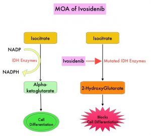
Isocitrate DeHydrogenase (IDH) is a metabolic enzyme that helps generate energy from glucose and other metabolites, by catalyzing the conversion of Isocitrate to Alpha-Ketoglutarate. Alpha-ketoglutarate is required to properly regulate DNA and histone methylation, which in turn is important for gene expression and cellular differentiation. IDH mutations lead to aberrant DNA methylation and altered gene expression thereby preventing cellular differentiation, with resulting immature undifferentiated cells. IDH mutations can thus promote leukemogenesis in Acute Myeloid Leukemia and tumorigenesis in solid tumors and can result in inferior outcomes. There are three isoforms of IDH. IDH1 is mainly found in the cytoplasm, as well as in peroxisomes, whereas IDH2 and IDH3 are found in the mitochondria, and are a part of the Krebs cycle. Approximately 20% of patients with AML, 70% of patients with Low-grade Glioma and secondary Glioblastoma, 50% of patients with Chondrosarcoma, 20% of patients with Intrahepatic cholangiocarcinoma, 30% of patients with Angioimmunoblastic T-cell lymphoma and 8% of patients with Myelodysplastic syndromes/Myeloproliferative neoplasms, are associated with IDH mutations.MOA-of-Ivosidenib

TIBSOVO® (Ivosidenib) is a first-in-class, oral, potent, targeted, small-molecule inhibitor of mutant IDH1. The FDA in 2018, approved TIBSOVO® for adult patients with relapsed or refractory AML with a susceptible IDH1 mutation and in 2019 approved TIBSOVO® for newly diagnosed AML with a susceptible IDH1 (Isocitrate DeHydrogenase-1) mutation, in patients who are at least 75 years old or who have comorbidities that preclude the use of intensive induction chemotherapy. VIDAZA® (Azacitidine) is a hypomethylating agent that promotes DNA hypomethylation by inhibiting DNA methyltransferases. VIDAZA® has been shown to significantly improve Overall Survival (OS) when compared to conventional care regimens in elderly unfit patients with newly diagnosed AML, who are not candidates for intensive chemotherapy. In a Phase Ib trial, TIBSOVO® in combination with VIDAZA® showed encouraging clinical activity in newly diagnosed IDH1-mutated AML patients.

AGILE is a global, double-blind, randomized, placebo-controlled, Phase III trial in which the efficacy and safety of a combination of TIBSOVO® and VIDAZA® were assessed, as compared with placebo and VIDAZA®, in patients with newly diagnosed IDH1-mutated Acute Myeloid Leukemia, who were ineligible for intensive induction chemotherapy. Patients were randomly assigned in a 1:1 ratio to receive TIBSOVO® 500 mg orally once daily combined with VIDAZA® 75 mg/m2 subcutaneously or IV for 7 days in 28-day cycles (N=72) or placebo and VIDAZA® (N=74). All the patients were to be treated for a minimum of six cycles until disease progression or unacceptable toxicities. The median patient age was 76 years, 75% had primary AML and 25% had secondary AML, 67% had intermediate cytogenetic risk and 22% had poor cytogenetic risk. Patients were stratified according to geographic region and disease status (Primary versus Secondary Acute Myeloid Leukemia). The Primary end point was Event-Free Survival, defined as the time from randomization until treatment failure (failure of complete remission by week 24), relapse from remission, or death from any cause, whichever occurred first.

At a median follow-up of 12.4 months, Event-Free Survival was significantly longer in the TIBSOVO® and VIDAZA® group than in the placebo and VIDAZA® group (HR=0.33; P=0.002). This benefit was seen across all key subgroups. The estimated probability that a patient would remain event-free at 12 months was 37% in the TIBSOVO® and VIDAZA® group and 12% in the placebo and VIDAZA® group. The median Overall Survival was 24.0 months with TIBSOVO® and VIDAZA® and 7.9 months with placebo and VIDAZA® (HR=0.44; P=0.001). Among those patients who were dependent on transfusion of red blood cells, platelets, or both at baseline, a higher percentage of patients converted to transfusion independence with TIBSOVO® and VIDAZA®, than with placebo and VIDAZA® (46% versus 18%; P=0.006). Health-Related Quality of Life scores favored TIBSOVO® and VIDAZA® across all subscales. Grade 3 or higher Adverse Events included febrile neutropenia (28% with TIBSOVO® and VIDAZA® versus 34% with placebo and VIDAZA®) and neutropenia (27% versus 16%, respectively). Differentiation syndrome of any grade occurred in 14% of the patients receiving TIBSOVO® and VIDAZA® versus 8% among those receiving placebo and VIDAZA®.

It was concluded that a combination of TIBSOVO® and VIDAZA® significantly improved Event-Free Survival, Response Rates, and Overall Survival, as compared with placebo and VIDAZA®, in patients with newly diagnosed IDH1-mutated Acute Myeloid Leukemia, who were ineligible for induction chemotherapy. The authors added that treatment with TIBSOVO® and VIDAZA® resulted in better Quality of Life and higher rates of transfusion independence.

Ivosidenib and Azacitidine in IDH1-Mutated Acute Myeloid Leukemia. Montesinos P, Recher C, Vives S, et al. N Engl J Med 2022; 386:1519-1531.

Overall Survival at 2 Years with LUMAKRAS® for KRAS G12C Positive Non Small Cell Lung Cancer

SUMMARY: The American Cancer Society estimates that for 2022, about 236,740 new cases of lung cancer will be diagnosed and 135,360 patients will die of the disease. Lung cancer is the leading cause of cancer-related mortality in the United States. Non-Small Cell Lung Cancer (NSCLC) accounts for approximately 85% of all lung cancers. Of the three main subtypes of NSCLC, 30% are Squamous Cell Carcinomas (SCC), 40% are Adenocarcinomas and 10% are Large Cell Carcinomas. With changes in the cigarette composition and decline in tobacco consumption over the past several decades, Adenocarcinoma now is the most frequent histologic subtype of lung cancer.

The KRAS (kirsten rat sarcoma viral oncogene homologue) proto-oncogene encodes a protein that is a member of the small GTPase super family. The KRAS gene provides instructions for making the KRAS protein, which is a part of a signaling pathway known as the RAS/MAPK pathway. By relaying signals from outside the cell to the cell nucleus, the protein instructs the cell to grow, divide and differentiate. The KRAS protein is a GTPase, and converts GTP into GDP. To transmit signals, the KRAS protein must be turned on, by binding to a molecule of GTP. When GTP is converted to GDP, the KRAS protein is turned off or inactivated, and when the KRAS protein is bound to GDP, it does not relay signals to the cell nucleus. The KRAS gene is in the Ras family of oncogenes, which also includes two other genes, HRAS and NRAS. When mutated, oncogenes have the potential to change normal cells cancerous.

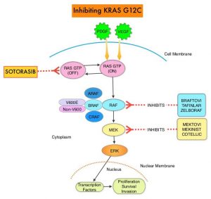
KRAS is the most frequently mutated oncogene in human cancers and are often associated with resistance to targeted therapies and poor outcomes. The KRAS-G12C mutation occurs in approximately 12-15% of Non Small Cell Lung Cancers (NSCLC) and in 3-5% of colorectal cancers and other solid cancers. KRAS G12C is one of the most prevalent driver mutations in NSCLC and accounts for a greater number of patients than those with ALK, ROS1, RET, and TRK 1/2/3 mutations combined. KRAS G12C cancers are genomically more heterogeneous and occur more frequently in current or former smokers, and are likely to be more complex genomically than EGFR mutant or ALK rearranged cancers. G12C is a single point mutation with a Glycine-to-Cysteine substitution at codon 12. This substitution favors the activated state of KRAS, resulting in a predominantly GTP-bound KRAS oncoprotein, amplifying signaling pathways that lead to oncogenesis.Inhibiting-KRAS-G12C

LUMAKRAS® (Sotorasib) is a first-in-class small molecule that specifically and irreversibly inhibits KRAS-G12C and traps KRAS-G12C in the inactive GDP-bound state. Preclinical studies in animal models showed that LUMAKRAS® inhibited nearly all detectable phosphorylation of Extracellular signal-Regulated Kinase (ERK), a key downstream effector of KRAS, leading to durable complete regression of KRAS-G12C tumors. The CodeBreaK clinical development program for LUMAKRAS® was designed to treat patients with an advanced solid tumor with the KRAS G12C mutation and address the longstanding unmet medical need for these cancers. This program has enrolled more than 800 patients across 13 tumor types since its inception.

CodeBreaK 100 is a Phase I and II, first-in-human, open-label, single arm, multicenter study, which enrolled patients with KRAS G12C-mutant solid tumors. Eligible patients must have received a prior line of systemic anticancer therapy, for their tumor type and stage of disease. The Phase II trial enrolled 126 patients with NSCLC, who had locally advanced or metastatic NSCLC with a KRAS G12C mutation, and had progressed on an immune checkpoint inhibitor and/or platinum-based chemotherapy. Patients with active brain metastases were excluded. Patient received LUMAKRAS® 960 mg orally once daily, until disease progression or unacceptable toxicity. The median age was 64 years, 52% were male, over 90% of patients had a smoking history, median number of prior lines of therapy was two, 92% had prior platinum-based chemotherapy and 90% had prior anti–PD-L1 therapy, 83% had both prior platinum-based chemotherapy and immunotherapy. The Primary end point of the trial was Overall Response Rate (ORR) as assessed by blinded Independent Central Review. Secondary end points included Duration of Response (DOR), Disease Control Rate (DCR), time to recovery, Progression Free Survival (PFS), Overall Survival (OS), and Safety. The examination of biomarkers served as an exploratory end point.

At the time of Primary analysis, at a median follow up of 12.2 months, the ORR was 37.1% and the median Duration of Response was 10 months. Based on the data from the primary analysis, the FDA in 2021 granted accelerated approval to LUMAKRAS®, for the treatment of patients with locally advanced or metastatic NSCLC, whose tumors harbor the KRAS G12C mutation, and who have received prior therapies.

For this updated analysis, the median follow up time for OS was 24.9 months, and the researchers included 174 patients enrolled in Phase I (N=48) and Phase II (N=126) portions of the CodeBreaK 100 trial, who were treated with LUMAKRAS®. The Overall Response Rate was 40.7% and the Disease Control Rate (DCR) was 83.7%. The median time to response was 6 weeks, the median Duration of Response was 12.3 month and 50.6% of responders remained in response for 12 months or more. The median PFS was 6.3 months and the median OS showed no change in the updated analysis, and was 12.5 months. At 1-year, the OS rate was 50.8% and the 2-year Overall Survival was 32.5%. The researchers performed additional analyses on both tumor and blood samples to identify biomarker profiles associated with durable clinical benefit and these showed that prolonged clinical benefit was observed regardless of Tumor Mutation Burden, PDL1 expression, and STK11 co-mutation status. Grade 3 or 4 treatment-related Adverse Events occurred in 21% of patients. Most adverse events were Grade 1 or 2, and treatment-related adverse events occurring in more than 10% of patients included diarrhea, elevated liver enzymes, nausea and fatigue.

It was concluded from this updated analysis that this is the longest follow up of patients on any KRAS G12C inhibitor, and LUMAKRAS® demonstrated meaningful and durable efficacy in patients with KRAS mutated NSCLC for whom treatment options are limited, following progression on first line treatment, and historically have had poor outcomes. Patients on LUMAKRAS® benefitted regardless of Tumor Mutation Burden, PDL1 expression, and STK11 co-mutation status. A global Phase III study (CodeBreaK 200) is underway, comparing LUMAKRAS® to Docetaxel in patients with KRAS G12C-mutated NSCLC.

Long-term outcomes with sotorasib in pretreated KRASp.G12C-mutated NSCLC: 2-year analysis of CodeBreaK100. Dy GK, Govindan R, Velcheti V, et al. Presented at: 2022 AACR Annual Meeting; April 8-13, 2022, New Orleans, LA. Abstract CT008.