Efficacy and Safety in the Phase 3 QUAZAR® AML-001 Study of ONUREG® (azacitidine) Tablets


Written by: Thomas E Boyd, MD, Texas Oncology
Content Sponsored by: Bristol Myers Squibb
Dr. Boyd is a paid consultant for BMS and was compensated for his contribution in drafting this article.


Acute myeloid leukemia (AML) is an aggressive hematologic cancer with 5-year relative survival rates less than 30%.1 The journey for patients diagnosed with AML is very complex, and helping patients achieve remission is a major goal of initial treatment.2 Relapse after this initial therapy poses a significant roadblock to survival, as evidenced by high relapse rates and the current 5-year survival rate.1,3 There remains a critical need for therapies that extend survival.4

After a patient enters first remission, continued AML treatment is one avenue for appropriate patients.4 ONUREG® is the first and only FDA-approved continued AML treatment for adult patients with acute myeloid leukemia who achieved first complete remission (CR) or complete remission with incomplete blood count recovery (CRi) following intensive induction chemotherapy and are not able to complete intensive curative therapy.5

The efficacy and safety of ONUREG® was evaluated in the Phase 3 QUAZAR®AML-001 trial, where patients who completed intensive induction with or without consolidation therapy were randomized 1:1 to receive ONUREG or placebo orally on Days 1 through 14 of each 28-day cycle.5 Randomization was stratified by age at time of induction therapy, cytogenetic risk category at time of induction therapy, prior history of MDS/CMML, and receipt of consolidation therapy following induction therapy.5

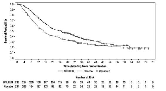
Median OS, the primary endpoint in the QUAZAR®AML-001 trial, was over 2 years (24.7 months) in the ONUREG® arm and 14.8 months in the placebo arm (hazard ratio [HR] 0.69, 95% confidence interval [CI]: 0.55-0.86; P=0.0009; Figure 1).5 This statistically significant survival benefit of ~10 months for patients with AML in first remission who received ONUREG® compared to placebo demonstrated that QUAZAR® AML-001 trial met its primary endpoint.5 Survival estimates were 73% for ONUREG® (n=168; 95% CI: 67, 78) and 56% for placebo (n=127; 95% CI: 49, 62) at 1 year, and 51% for ONUREG® (n=115; 95% CI: 44, 57) and 37% for placebo (n=82; 95% CI: 31, 43) at 2 years.6 Analyses of survival estimates at 1 and 2 years were not designed to show a difference between treatment arms.

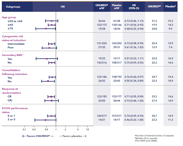
Additional analyses show the influence of ONUREG® across subgroups vs placebo for median OS (Figure 2). It is important to note that these prespecified analyses should be interpreted with caution because of potential selection bias, insufficient sample size, and a higher probability of making a false positive finding.7

The most common adverse reactions (ARs, ≥ 10%) associated with ONUREG® treatment included nausea, vomiting, diarrhea, fatigue/asthenia, constipation, pneumonia, abdominal pain, arthralgia, decreased appetite, febrile neutropenia, dizziness, and pain in extremity.5 Serious ARs occurred in 15% of patients who received ONUREG®, and the most common Grade 3/4 ARs were febrile neutropenia (11%), pneumonia (9%), diarrhea (5%), and fatigue/asthenia (4%).5

Efficacy and safety were established by the large, multicenter QUAZAR® AML-001 trial. These results established ONUREG® as a continued treatment for adult patients with AML who achieve first complete remission (CR) or complete remission with incomplete blood count recovery (CRi) following intensive induction chemotherapy and are not able to complete intensive curative therapy.

Figure 1: Kaplan-Meier curve for OS: ITT population in QUAZAR® AML-0015
KM-for-Overall-Survival


Figure 2: Overall survival select subgroup analysis6,7
Overall-Survival


*Analysis limitations: These prespecified subgroup analyses should be interpreted with caution to determine a difference between arms in these select subgroups because of potential selection bias, insufficient sample size, and a higher probability of making a false positive finding.

IMPORTANT SAFETY INFORMATION
CONTRAINDICATIONS
ONUREG® is contraindicated in patients with known severe hypersensitivity to azacitidine or its components.
WARNINGS AND PRECAUTIONS
Risks of Substitution with Other Azacitidine Products
Due to substantial differences in the pharmacokinetic parameters, the recommended dose and schedule for ONUREG® are different from those for the intravenous or subcutaneous azacitidine products. Treatment of patients using intravenous or subcutaneous azacitidine at the recommended dosage of ONUREG® may result in a fatal adverse reaction. Treatment with ONUREG® at the doses recommended for intravenous or subcutaneous azacitidine may not be effective. Do not substitute ONUREG® for intravenous or subcutaneous azacitidine.
Myelosuppression
New or worsening Grade 3 or 4 neutropenia and thrombocytopenia occurred in 49% and 22% of patients who received ONUREG®. Febrile neutropenia occurred in 12%. A dose reduction was required for 7% and 2% of patients due to neutropenia and thrombocytopenia. Less than 1% of patients discontinued ONUREG® due to either neutropenia or thrombocytopenia. Monitor complete blood counts and modify the dosage as recommended. Provide standard supportive care, including hematopoietic growth factors, if myelosuppression occurs.
Increased Early Mortality in Patients with Myelodysplastic Syndromes (MDS)
In AZA-MDS-003, 216 patients with red blood cell transfusion-dependent anemia and thrombocytopenia due to MDS were randomized to ONUREG® or placebo. 107 received a median of 5 cycles of ONUREG® 300 mg daily for 21 days of a 28-day cycle. Enrollment was discontinued early due to a higher incidence of early fatal and/or serious adverse reactions in the ONUREG® arm compared with placebo. The most frequent fatal adverse reaction was sepsis. Safety and effectiveness of ONUREG® for MDS have not been established. Treatment of MDS with ONUREG® is not recommended outside of controlled trials.
Embryo-Fetal Toxicity
ONUREG® can cause fetal harm when administered to a pregnant woman. Azacitidine caused fetal death and anomalies in pregnant rats via a single intraperitoneal dose less than the recommended human daily dose of oral azacitidine on a mg/m2 basis. Advise pregnant women of the potential risk to a fetus. Advise females of reproductive potential to use effective contraception during treatment with ONUREG® and for at least 6 months after the last dose. Advise males with female partners of reproductive potential to use effective contraception during treatment with ONUREG® and for at least 3 months after the last dose.
ADVERSE REACTIONS
Serious adverse reactions occurred in 15% of patients who received ONUREG®. Serious adverse reactions in ≥2% included pneumonia (8%) and febrile neutropenia (7%). One fatal adverse reaction (sepsis) occurred in a patient who received ONUREG®.
Most common (≥10%) adverse reactions with ONUREG® vs placebo were nausea (65%, 24%), vomiting (60%, 10%), diarrhea (50%, 21%), fatigue/asthenia (44%, 25%), constipation (39%, 24%), pneumonia (27%, 17%), abdominal pain (22%, 13%), arthralgia (14%, 10%), decreased appetite (13%, 6%), febrile neutropenia (12%, 8%), dizziness (11%, 9%), pain in extremity (11%, 5%).
LACTATION
There are no data regarding the presence of azacitidine in human milk or the effects on the breastfed child or milk production. Because of the potential for serious adverse reactions in the breastfed child, advise women not to breastfeed during treatment with ONUREG® and for 1 week after the last dose.

Please see full Prescribing Information for ONUREG®.

References
1. National Cancer Institute. SEER Cancer Statistics Factsheets: Acute Myeloid Leukemia. http://seer.cancer.gov/statfacts/html/amyl.html. Accessed April 21, 2021.
2. Medeiros BC. Interpretation of clinical endpoints in trials of acute myeloid leukemia. Leuk Res. 2018;68:32-29.
3. Wei AH. Maintenance therapy for AML: are we there yet? Blood. 2019;133(13):1390-1392.
4. Medeiros BC, Chan SM, Daver NG, Jonas BA, Pollyea DA. Optimizing survival outcomes with post-remission therapy in acute myeloid leukemia. Am J Hematol. 2019;94:803-811.
5. ONUREG® [Prescribing Information]. Summit, NJ: Celgene Corporation; 2021.
6. Wei AH, Döhner H, Pocock C, et al. The QUAZAR AML-001 maintenance trial: results of a phase III international, randomized, double-blind, placebo-controlled study of CC-486 in patients with acute myeloid leukemia (AML) in first remission [oral presentation at ASH 2019]. Blood. 2019;134(Suppl 2):LBA-3.
7. Wei AH, Döhner H, Pocock C, et al. Oral Azacitidine Maintenance Therapy for Acute Myeloid Leukemia in First Remission. N Engl J Med. 2020;383:2526-2537.

© 2021 Celgene Corporation.
ONUREG is a trademark of Celgene Corporation, a Bristol Myers Squibb company.
QUAZAR® is a registered trademark of Celgene Corporation, a Bristol Myers Squibb company
02/22 2011-US-2100200

Association of Gut Microbiome with Immune Checkpoint Inhibitor Response in Advanced Melanoma

SUMMARY: The American Cancer Society estimates that in 2022, there will be an estimated 1.92 million new cancer cases diagnosed and 609,360 cancer deaths in the United States. Immunotherapy with Immune Checkpoint Inhibitors (ICIs) has revolutionized cancer care and has become one of the most effective treatment options by improving Overall Response Rate and prolongation of survival across multiple tumor types. These agents target Programmed cell Death protein-1 (PD-1), Programmed cell Death Ligand-1 (PD-L1), Cytotoxic T-Lymphocyte-Associated protein-4 (CTLA-4), and many other important regulators of the immune system. Over 50% of patients treated with a combination of PD-1 and CTLA-4 inhibitors are alive after five years. Nonetheless, less than 50% of the patients respond to single-agent ICI and a higher response to targeting both PD-1 and CTLA-4 is associated with significant immune-related Adverse Events.

Biomarkers predicting responses to ICI’s include Tumor Mutational Burden (TMB), Mismatch Repair (MMR) status, and Programmed cell Death Ligand 1 (PD‐L1) expression. Other biomarkers such as Tumor Infiltrating Lymphocytes (TILs), TIL‐derived Interferon‐γ, Neutrophil‐to‐Lymphocyte ratio, and peripheral cytokines, have also been proposed as predictors of response. It has been postulated that concomitant medications during therapy with ICIs such as baseline steroid use as well as treatment with antibiotics may negate or lessen the efficacy of ICIs.

Preclinical studies have suggested that immune-based therapies for cancer may have a very complex interplay with the host’s microbiome and there may be a relationship between gut bacteria and immune response to cancer. The gut microbiome is unique in each individual, including identical twins. The crosstalk between microbiota in the gut and the immune system allows for the tolerance of commensal bacteria (normal microflora) and oral food antigens and at the same time enables the immune system to recognize and attack opportunistic bacteria. Immune Checkpoint Inhibitors strongly rely on the influence of the host’s microbiome, and the gut microbial diversity enhances mucosal immunity, dendritic cell function, and antigen presentation. Broad-spectrum antibiotics can potentially alter the bacterial composition and diversity of our gut microbiota, by killing the good bacteria. It has been postulated that this may negate the benefits of immunotherapy and influence treatment outcomes. It should be noted however that the relationship between gut bacteria and immune response is influenced by several factors and may be partially cancer type specific and it is unlikely that the same microbiome features can reflect the uniqueness of the genetic and immune characteristics of each tumor.

Even though the composition of the gut microbiome has been associated with clinical responses to immune checkpoint inhibitor (ICI) treatment, there is a lack of consistency of results between the published studies, and there is limited consensus on the specific microbiome characteristics linked to the clinical benefits of ICIs. The Predicting Response to Immunotherapy for Melanona with Gut Microbiome and Metabolomics (PRIMM) studies are two separate prospective observational cohort studies that has been recruiting patients in the UK (PRIMM-UK) and the Netherlands (PRIMM-NL) since 2018. These cohorts of previously ICI-naive patients with advanced melanoma have provided extensive biosamples, including stool, serum and peripheral blood mononuclear cells, before and during ICI treatment, with detailed clinical and dietary data collected at regular intervals longitudinally.

The authors therefore performed a meta-analysis on existing publicly available datasets to produce the largest study to date. In order to study the role of the gut microbiome in ICI response, the researchers recruited ICI-naive patients with advanced cutaneous melanoma from the PRIMM cohorts, as well as three additional cohorts of ICI-naive patients with advanced cutaneous melanoma, originating from Barcelona, Leeds and Manchester (N = 165), and performed shotgun metagenomic sequencing on a total of 165 stool microbiome samples collected before initiating ICI treatment. Shotgun sequencing is a laboratory technique for determining the DNA sequence of an organism’s genome. This dataset was integrated with 147 metagenomic samples from smaller publicly available datasets. This methodology provided the largest assessment of the potential of the gut microbiome as a biomarker of response to ICI, in addition to allowing for investigation of specific microbial species or functions associated with response. Patient demographics including age, gender, BMI, previous non-immunotherapy treatments, previous drug therapies such as antibiotics, Proton Pump Inhibitors (PPIs) and steroids, as well as dietary patterns, were collected in these cohorts for the majority of patients, and were considered in the multivariate analysis.

The researchers used machine learning analysis to understand the association between gut microbiome and response to ICIs. This analysis confirmed the link between the microbiome and Overall Response Rates (ORRs), as well as Progression Free Survival (PFS) with ICIs. This analysis also revealed limited reproducibility of microbiome-based signatures across cohorts. A panel of species, including Bifidobacterium pseudocatenulatum, Roseburia spp. and Akkermansiamuciniphila were associated with responders, but no single species could be regarded as a fully reliable biomarker across studies. Based on these findings from this large set of real-world cohorts, the authors noted that the relationship between human gut microbiome and response to ICIs is more complex than previously understood, and extends beyond the presence or absence of different microbial species in responders and nonresponders.

It was concluded that future studies should include large samples and take into account the complex interplay of clinical factors with the gut microbiome over the treatment course. Until then, the authors recommend high-quality, diverse, whole-foods diet to optimize gut health, rather than consumption of commercial probiotics.

Cross-cohort gut microbiome associations with immune checkpoint inhibitor response in advanced melanoma. Lee KA, Thomas AM, Bolte LA, et al. Nat Med. 2022 Feb 28. doi: 10.1038/s41591-022-01695-5. Online ahead of print.

Avoiding Radioiodine Therapy following Thyroidectomy in Patients with Low-Risk Thyroid Cancer

SUMMARY: The American Cancer Society estimates that about 43,800 new cases of thyroid cancer will be diagnosed in the United States in 2022 and about 2,230 patients will die of the disease. Differentiated Thyroid Cancer (DTC) is the most common endocrine malignancy and includes Papillary, Follicular, and Hürthle-cell cancers, with Papillary thyroid cancers accounting for 80% of them. Majority of patients with DTC have clinical Stage I or Stage II disease, with a recurrence rate of less than 5% and cancer-related death rates even lower. Risk factors for recurrence include tumor size, multifocality, capsular or angioinvasion, degree of cervical lymph node involvement, existence of BRAF V600E Mutation, and thyroglobulin levels more than 0.5 ng/mL, after thyroidectomy.

Even though Radioiodine (iodine-131) therapy is not recommended for patients with a unifocal microcarcinoma (10 mm or less in diameter) following thyroidectomy, Radioiodine therapy is generally offered to a majority of patients with low-risk thyroid cancer, both to ablate residual normal thyroid tissue and to treat unresectable persistent disease. The benefits of this intervention however remain controversial. .

The authors conducted a prospective, multicenter, randomized, Phase III Essai Stimulation Ablation 2 (ESTIMABL2) trial involving patients with low-risk thyroid cancer, to assess the non-inferiority of observation versus postoperative Radioiodine therapy, following thyroidectomy. In this study, a total of 776 patients with low-risk Differentiated Thyroid Cancer who were undergoing thyroidectomy were randomly assigned 1:1 to receive ablation with postoperative Radioiodine therapy at a dose of 1.1 GBq (N=389) or no Radioiodine therapy (N=387). Enrolled patients had Differentiated Thyroid Carcinoma (Papillary, Follicular, or Oncocytic/Hürthle-cell cancer), with a multifocal pT1a tumor or a pT1b tumor. None of the patients had regional lymph node involvement, extrathyroidal extension or aggressive histologic subtypes (tall-cell, clear-cell, columnar-cell, and diffuse sclerosing variants of Papillary thyroid cancer, poorly differentiated). The mean patient age was 52 years, and 83% were women, 96% had papillary tumors 81% had pT1b N0 or Nx disease. All patients had normal results on postoperative neck ultrasonography. The follow-up protocol consisted of the measurement of thyroglobulin and thyroglobulin antibodies in all patients at 10 months and yearly thereafter. Ultrasonography of the neck was performed in all patients 10 months and 3 years after randomization. Disease-related events included residual or recurrent disease on neck ultrasonography and a serum thyroglobulin level of more than 1 ng/mL in the group receiving radioiodine and a level of more than 5 ng/mL in the nontreated group. No diagnostic Radioiodine scanning was performed after the whole-body scanning that was performed after therapy. The Primary objective was to assess whether no Radioiodine therapy was noninferior to Radioiodine therapy, with respect to the absence of a composite end point that included functional, structural, and biologic abnormalities, indicating residual or recurrent disease at 3 years.

After 3 years of follow up, there were no clinically meaningful differences in any of the end points between the two groups and the percentage of patients without an event was 95.6% in the no-Radioiodine therapy group and 95.9% in the Radioiodine therapy group, a result that met the noninferiority criteria. Events were more frequent in patients with a postoperative serum thyroglobulin level of more than 1 ng/mL during thyroid hormone treatment. BRAF V600E molecular alterations, which are associated aggressive tumor characteristics, were found in approximately 50% of the samples in each treatment group. The mutational status did not influence event rates in these low-risk patients. No treatment-related adverse events were reported and there was no difference in Quality-of-Life scores between the two groups.

It was concluded that in patients with low-risk thyroid cancer undergoing thyroidectomy, follow up without the use of Radioiodine therapy was noninferior to an ablation strategy with Radioiodine therapy, suggesting that patients with low-risk disease generally do well regardless of whether they receive Radioiodine therapy.

Thyroidectomy without Radioiodine in Patients with Low-Risk Thyroid Cancer. Leboulleux S, Bournaud C, Chougnet CN, et al. N Engl J Med 2022; 386:923-932

PET Response-Based Radiotherapy De-Escalation in p16 Positive Oropharyngeal Cancer

SUMMARY: The American Cancer Society estimates that about 54,000 new cases of oral cavity or Oropharyngeal Cancer will be diagnosed in the US in 2022 and about 11,230 patients will die of the disease. According to the CDC, based on data from 2014 to 2018, about 46,143 HPV-associated cancers occur in the United States each year (25,719 among women, and 20,424 among men). Cervical cancer is the most common HPV-associated cancer among women, and Oropharyngeal cancers are the most common among men. There has been a significant increase in the incidence during the past several decades, due to changes in sexual practices.

HPV-positive Oropharyngeal Squamous Cell Carcinoma (OPSCC) is an entirely distinct disease entity from HPV-negative Oropharyngeal Squamous Cell Carcinoma. Patients with HPV-positive OPSCC tend to be younger males, who are former smokers or nonsmokers, with risk factors for exposure to High Risk HPV (HR-HPV). The HPV-positive primary Squamous Cell Carcinoma tend to be smaller in size, with early nodal metastases, and these patients have a better prognosis compared with patients with HPV-negative Head and Neck Squamous Cell Carcinoma (HNSCC), when treated similarly. Expression of tumor suppressor protein, known as p16, is highly correlated with infection with HPV in HNSCC. Accurate HPV assessment in head and neck cancers is becoming important as it significantly impacts clinical management. HPV status is considered the most important prognostic indicator in patients with head and neck cancer and p16 status is now included in the American Joint Committee on Cancer (AJCC) Staging System.

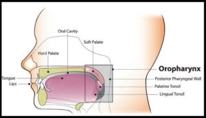
HPV-positive Oropharyngeal Squamous Cell Carcinoma is more sensitive to chemotherapy and radiotherapy than is HPV-negative Oropharyngeal Squamous Cell Carcinoma, which translates to a much better prognosis and survival, when treated with a combination of Cisplatin chemotherapy and Radiotherapy. This treatment however can be associated with substantial morbidity and lifelong toxicities such as dry mouth, difficulty swallowing, and loss of taste.Oropharynx

To address these toxicities, the authors conducted a prospective Phase II de-escalation study utilizing FDG-PET response criteria to select patients eligible for de-escalated radiotherapy. The researchers hypothesized that early swallow function and quality of life will improve with lower dose radiation. This study enrolled 59 patients (N=59) with Stage I-II (AJCC) p16-positive Oropharyngeal cancer, with FDG-avid disease and any smoking history. Patients with matted lymph nodes or history of head and neck surgery were excluded.

All enrolled patients had a pre-treatment FDG-PET/CT imaging and received weekly Carboplatin and Paclitaxel concurrently with planned radiotherapy at 70 Gy in 35 fractions. FDG-PET was repeated midway through treatment after 2 weeks at fraction 10. Patients who had tumors with lower metabolic activity before treatment and more than 50% reduction in Metabolic Tumor Volume after 2 weeks of treatment were de-escalated from the standard radiotherapy total dose of 70 Gy in 35 fractions, to a total dose of 54 Gy in 27 fractions. The median patient age was 60 years and baseline characteristics showed that both standard and de-escalated cohorts had similar patient demographics and pathology. At the planned interim analysis, early toxicity and Patient Reported Outcomes (PROs) were examined.

Fifty percent (50%) of the patients met de-escalation criteria and received the lower radiation dose, leading to 20-30% reductions in radiation exposure to sensitive structures in the head and neck prone to toxicities such as larynx, constrictors, oral cavity and salivary glands. De-escalation from the standard radiotherapy resulted in significantly less acute toxicity, and at one month after treatment, patients who received de-escalated therapy lost less weight as a percentage of baseline, compared to standard treatment group (6% versus 11%; P<0.001) and had improved videofluoroscopic swallowing function following treatment, and fewer patients required feeding tube placement during treatment.

The authors concluded that although the trial remains ongoing, mid-treatment FDG-PET response adaptation allows for approximately 50% of early stage p16 positive Oropharyngeal cancer patients to be de-escalated to a total dose of 54 Gy. This in turn can result in approximately 25% reduction in dose delivered to organs known to affect toxicity and quality of life, with significantly better objective measures of toxicity and numerically improved Patient Related Outcomes.

Early Toxicity and Patient Reported Outcomes From a Phase 2 Trial of FDG-PET Response-Based De-Escalated Definitive Radiotherapy for p16+ Oropharynx Cancer. Allen SG, Rosen BS, Aryal MP, et al. 2022 Multidisciplinary Head and Neck Symposium; February 24-26, 2022; Phoenix, AZ. Abstract 1.

LYNPARZA® Superior to Next-Generation Hormonal Drug in CRPC Patients with Homologous Recombination Repair Gene Alterations

SUMMARY: Prostate cancer is the most common cancer in American men with the exclusion of skin cancer, and 1 in 9 men will be diagnosed with Prostate cancer during their lifetime. It is estimated that in the United States, about 268,490 new cases of Prostate cancer will be diagnosed in 2022 and 34,500 men will die of the disease. The development and progression of Prostate cancer is driven by androgens. Androgen Deprivation Therapy (ADT) or testosterone suppression has therefore been the cornerstone of treatment of advanced Prostate cancer and is the first treatment intervention. Approximately 10-20% of patients with advanced Prostate cancer will progress to Castration Resistant Prostate Cancer (CRPC) within five years during ADT, and over 80% of these patients will have metastatic disease at the time of CRPC diagnosis. The malignant transformation of prostatic epithelial cell as well as the development of CRPC has been attributed to deleterious alterations in a variety of genes including loss-of-function alterations in Homologous Recombination Repair (HRR) genes.

DNA damage is a common occurrence in daily life by UV light, ionizing radiation, replication errors, chemical agents, etc. This can result in single and double strand breaks in the DNA structure which must be repaired for cell survival. The two vital pathways for DNA repair in a normal cell are BRCA1/BRCA2 and PARP. BRCA1 and BRCA2 are tumor suppressor genes that recognize and repair double strand DNA breaks via Homologous Recombination Repair (HRR) pathway. Homologous Recombination is a type of genetic recombination, and is a DNA repair pathway utilized by cells to accurately repair DNA double-stranded breaks during the S and G2 phases of the cell cycle, and thereby maintain genomic integrity. Homologous Recombination Deficiency (HRD) is noted following mutation of genes involved in HR repair pathway. At least 15 genes are involved in the Homologous Recombination Repair (HRR) pathway including BRCA1, BRCA2, PALB2, CHEK2 and ATM genes. Mutations in these genes predispose an individual to develop malignant tumors. Mutations in BRCA1 and BRCA2 account for about 20-25% of hereditary breast cancers and about 5-10% of all breast cancers. They also account for 15% of ovarian cancers, in addition to other cancers such as Colon and Prostate. BRCA mutations can either be inherited (Germline) and present in all individual cells or can be acquired and occur exclusively in the tumor cells (Somatic). Somatic mutations account for a significant portion of overall BRCA1 and BRCA2 aberrations. Loss of BRCA function due to frequent somatic aberrations likely deregulates HR pathway, and other pathways then come in to play, which are less precise and error prone, resulting in the accumulation of additional mutations and chromosomal instability in the cell, with subsequent malignant transformation. Homologous Recombination Deficiency therefore indicates an important loss of DNA repair function.

The PARP (Poly ADP Ribose Polymerase), family of enzymes include, PARP1 and PARP2, and is a related enzymatic pathway that repairs single strand breaks in DNA. In a BRCA mutant, the cancer cell relies solely on PARP pathway for DNA repair to survive. PARP inhibitors trap PARP onto DNA at sites of single-strand breaks, preventing their repair and generating double-strand breaks that cannot be repaired accurately in tumors harboring defects in Homologous Recombination Repair pathway genes, such as BRCA1 or BRCA2 mutations, and this leads to cumulative DNA damage and tumor cell death.

LYNPARZA® (Olaparib) is a first-in-class PARP inhibitor and blocks DNA damage response in tumors harboring a deficiency in Homologous Recombination Repair, as is noted in those with mutations such as BRCA1 and/or BRCA2. LYNPARZA® showed promising results in a Phase II trial (TOPARP), when given as monotherapy, in patients with BRCA1/2 or ATM gene-mutated mCRPC, who had received a prior Taxane-based chemotherapy, and at least one newer hormonal agent (ZYTIGA® or XTANDI®).

PROfound is a prospective, multicentre, randomized, open-label, Phase III trial in which the efficacy and safety of LYNPARZA® was compared with physician’s choice of either XTANDI® or ZYTIGA® in two groups of patients with mCRPC, who had progressed on prior treatment with new hormonal anticancer treatments, and had a qualifying tumor mutation in one of 15 genes involved in the Homologous Recombination Repair (HRR) pathway. Patients in Cohort A (N=245) had alterations in BRCA1, BRCA2 or ATM genes while those in Cohort B (N=142) had alterations in any one of 12 other genes known to be involved in DNA repair which included BRIP1, BARD1, CDK12, CHEK1, CHEK2, FANCL, PALB2, PPP2R2A, RAD51B, RAD51C, RAD51D or RAD54L. Patients were randomized 2:1 within each cohort to receive LYNPARZA® 300 mg orally BID or physician’s choice of XTANDI® 160 mg orally QD or ZYTIGA® 1000 mg orally QD along with Prednisone 5 mg orally BID. Patient characteristics were well-balanced between arms in each treatment group, median patient age was 68 years, approximately 25% of patients had de novo metastatic disease, about 65% of patients received prior Taxane therapy and more than 20% had received two lines of chemotherapy. Patients were allowed to cross over to LYNPARZA® upon progression. The Primary endpoint was radiographic Progression-Free Survival (rPFS) in Cohort A, assessed by Blinded Independent Central Review (BICR).

The authors had previously reported that in Cohort A, the median PFS was 7.4 months with LYNPARZA®, compared to 3.5 months in the control group (HR=0.34, P<0.0001). This represented a 66% greater delay in disease progression compared to hormonal therapy. The interim Overall Survival analysis in Cohort A showed that median OS was 18.5 months with LYNPARZA® compared to 15 months with control drug treatment (HR=0.64, P=0.0173). In Cohort A, the Objective Response Rate (ORR) was 33.3% with LYNPARZA® compared with 2.3% with control drug therapies (P<0.0001).

The authors in this publication reported the results of the prespecified Secondary endpoints, which included pain, Health-Related Quality of Life (HRQOL), symptomatic Skeletal-Related Events, and time to first opiate use for cancer-related pain in Cohort A group of patients. Pain was assessed with the Brief Pain Inventory-Short Form, and HRQOL was assessed with the Functional Assessment of Cancer Therapy-Prostate (FACT-P). Cohort A included 245 patients with alterations in BRCA1, BRCA2, or ATM genes, of whom 162 patients received the investigational agent LYNPARZA®, and 83 patients received control drug. The median duration of follow up at data cutoff was 6.2 months for all LYNPARZA® group patients and 3.5 months for the control group patients.

The median time to pain progression was significantly longer with LYNPARZA® and was Not Reached in the LYNPARZA® group versus 9.92 months in the control group (HR=0.44; P=0.019). Pain interference scores were also significantly better in the LYNPARZA® group (difference in overall adjusted mean change from baseline score −0.85; nominal P=0.0004). Median time to progression of pain severity was Not Reached in either group. Among patients who had not used opiates at baseline (113 in the LYNPARZA® group, 58 in the control group), median time to first opiate use for cancer-related pain was 18.0 months in the LYNPARZA® group versus 7.5 months in the control group (HR=0.61; nominal P=0.044).

The proportion of patients with clinically meaningful improvement in FACT-P total score during treatment was higher for the LYNPARZA® group than the control group (10% versus 1% respectively; odds ratio=8.32; nominal P=0.0065). The median time to first symptomatic Skeletal-Related Event was not reached for either treatment group and the proportions of patients remaining free of symptomatic Skeletal-Related Events were 89.5% versus 77.1% at 6 months and 77.6% versus 53.6% at 12 months, in the LYNPARZA® and control groups respectively.

It was concluded that LYNPARZA® was associated with reduced pain burden and better-preserved HRQOL compared with the two control drugs, in patients with metastatic Castration-Resistant Prostate Cancer and Homologous Recombination Repair gene alterations, who had disease progression after a previous next-generation hormonal drug. The authors added that the study findings support the clinical benefit of improved radiographical Progression Free Survival and Overall Survival identified in PROfound trial.

Pain and health-related quality of life with olaparib versus physician’s choice of next-generation hormonal drug in patients with metastatic castration-resistant prostate cancer with homologous recombination repair gene alterations (PROfound): an open-label, randomised, phase 3 trial. Thiery-Vuillemin A, de Bono J, Hussain M, et al. Published:February 11, 2022. DOI:https://doi.org/10.1016/S1470-2045(22)00017-1

NUBEQA® Combination Improves Overall Survival in Metastatic Hormone Sensitive Prostate Cancer

SUMMARY: Prostate cancer is the most common cancer in American men with the exclusion of skin cancer, and 1 in 9 men will be diagnosed with Prostate cancer during their lifetime. It is estimated that in the United States, about 268,490 new cases of Prostate cancer will be diagnosed in 2022 and 34,500 men will die of the disease. The development and progression of Prostate cancer is driven by androgens. Androgen Deprivation Therapy (ADT) or testosterone suppression has therefore been the cornerstone of treatment of advanced Prostate cancer and is the first treatment intervention.

The first-generation NonSteroidal Anti-Androgen (NSAA) agents such as EULEXIN® (Flutamide), CASODEX® (Bicalutamide) and NILANDRON® (Nilutamide) act by binding to the Androgen Receptor (AR) and prevent the activation of the AR and subsequent up-regulation of androgen responsive genes. They may also accelerate the degradation of the AR. These agents have a range of pharmacologic activity from being pure anti-androgens to androgen agonists. CASODEX® is often prescribed along with GnRH (Gonadotropin-Releasing Hormone) agonists for metastatic disease, or as a single agent second line hormonal therapy for those who had progressed on LHRH agonists.

NUBEQA® (Darolutamide) is a potent second-generation Androgen Receptor (AR) inhibitor with a new chemical structure and has a high affinity to the AR. NUBEQA® does not cross the blood-brain barrier and for this reason has a favorable safety and tolerability profile in prespecified adverse events such as seizures, when compared with other second-generation AR inhibitors such as ERLEADA® (Apalutamide) and XTANDI® (Enzalutamide). It has been associated with increased Overall Survival (OS) among patients with non-metastatic Castration-Resistant Prostate Cancer (CRPC) and has been approved by the FDA for this indication. Whether a combination of NUBEQA®, in combination with Androgen Deprivation Therapy (ADT), and Docetaxel would increase survival among patients with metastatic Hormone-Sensitive Prostate Cancer, is unknown.

ARASENS is an international, randomized, double-blind, placebo-controlled, Phase III trial, which evaluated the efficacy and safety of NUBEQA® (Darolutamide) added to Androgen Deprivation Therapy (ADT) and Docetaxel in patients with metastatic Hormone Sensitive Prostate Cancer. In this study, a total of 1306 patients were randomly assigned 1:1 to receive NUBEQA® (N=651) or placebo (N=655), both in combination with ADT and Docetaxel. All the patients received ADT (either a Luteinizing Hormone Releasing Hormone (LHRH} agonist or antagonist) or underwent Orchiectomy within 12 weeks before randomization and received six cycles of Docetaxel 75 mg/m2 IV given on Day 1 every 21 days, with Prednisone or Prednisolone. Patients received LHRH agonists along with a first-generation anti-androgen agent for at least 4 weeks before randomization to help prevent a tumor flare, and the anti-androgen agent was discontinued before randomization. Patients were then randomly assigned to receive either NUBEQA® 600 mg orally twice daily or matched placebo, and treatment was continued until disease progression or unacceptable toxicities.

Eligible patients had biopsy proven prostate cancer with bone metastases and had to be candidates for ADT and Docetaxel. Patients with regional lymph node involvement only (N1, below the aortic bifurcation) or if they had received ADT more than 12 weeks before randomization, second-generation Androgen Receptor pathway inhibitors, chemotherapy, or immunotherapy for prostate cancer before randomization, or radiotherapy within 2 weeks before randomization, were excluded. The median age was 67 years and both treatment groups were well balanced. All patients had metastatic disease at baseline, 78% of the patients had a Gleason score of 8 or greater, about 80% had bone metastases (Stage M1b) and 18% had visceral metastases (Stage M1c). The Primary end point was Overall Survival (OS) and Secondary end points included were time to Castration-Resistant Prostate Cancer, time to pain progression, symptomatic Skeletal Event-Free Survival and time to initiation of subsequent systemic antineoplastic therapy, as well as Safety. The median follow up for Overall Survival was 43 months.

The median Overall Survival was not estimable in the NUBEQA® group versus 48.9 months in the placebo group. The addition of NUBEQA® to the combination with ADT and Docetaxel reduced the risk of death by 32%, compared to the placebo group (HR=0.68; P<0.001). This OS benefit was noted across most subgroups. Further, the significant OS benefit with the addition of NUBEQA® was observed, despite receipt of subsequent life-prolonging systemic therapies such as different Androgen-Receptor pathway inhibitors by 75.6% of patients in the placebo control group. The OS at 4 years was 62.7% in the NUBEQA® group and 50.4% in the placebo group.

With regard to Secondary endpoints, the addition of NUBEQA® to ADT and Docetaxel demonstrated consistent benefits. The time to development of Castration-Resistant Prostate Cancer was significantly longer in the NUBEQA® group (HR=0.36; P<0.001), the time to pain progression was also significantly longer in the NUBEQA® group (HR=0.79; P=0.01), as well as symptomatic Skeletal Event-Free Survival (HR=0.61; P<0.001). Further, the time to the initiation of subsequent systemic antineoplastic therapy was also significantly longer in the NUBEQA® group (HR=0.39; P<0.001). Adverse events were similar in the two groups.

The authors concluded that among patients with metastatic Hormone Sensitive Prostate Cancer, the addition of NUBEQA® to Androgen Deprivation Therapy and Docetaxel resulted in significantly longer Overall Survival, as well as improvement in key Secondary end points, with no increase in adverse events.

Darolutamide and Survival in Metastatic, Hormone-Sensitive Prostate Cancer. Smith MR, Hussain Saad F, et al. for the ARASENS Trial Investigators. NEJM. February 17, 2022. DOI: 10.1056/NEJMoa2119115.

Smoking Cessation after Lung Cancer Diagnosis Improves Overall Survival

SUMMARY: According to the American Cancer Society, tobacco use is responsible for about 1 in 5 deaths in the United States and is the leading preventable cause of death in the US. Smoking (cigarettes, cigars, and pipes) is responsible for about 20% of all cancers and about 30% of all cancer deaths in the US. Approximately 80% of lung cancers, as well as about 80% of all lung cancer deaths, are due to smoking, and lung cancer is the leading cause of cancer death in both men and women. Smoking also increases the risk for cancers of the Oral cavity, Oropharynx, Larynx, Esophagus, Stomach, Liver, Pancreas, Colon/Rectum, Kidney, Bladder, Cervix, as well as Acute Myeloid Leukemia. The American Cancer Society estimates that for 2022, about 236,740 new cases of lung cancer will be diagnosed and 135,360 patients will die of the disease. Non-Small Cell Lung Cancer (NSCLC) accounts for approximately 85% of all lung cancers. Of the three main subtypes of NSCLC, 30% are Squamous Cell Carcinomas (SCC), 40% are Adenocarcinomas and 10% are Large Cell Carcinomas. With changes in the cigarette composition and decline in tobacco consumption over the past several decades, Adenocarcinoma now is the most frequent histologic subtype of lung cancer.

Previous published studies have shown that individuals who start smoking at a younger age have greater mortality risk than those who start smoking later in life, and smoking cessation especially at younger ages substantially reduces that mortality risk. Several biologic mechanisms have been hypothesized, to explain the beneficial effect of smoking cessation on survival, in patients with Lung Cancer. Tobacco smoke has been shown to promote tumor growth, progression, and dissemination, while decreasing the efficacy and tolerance to radiation and systemic therapy. Further, it is well established that smoking increases the risk of postoperative complications and second primary cancers. Epigenetic changes induced by tobacco smoke may play an important role, and cigarette smoke induced diseases may be the result of alterations in DNA methylation, and is a reversible gene regulatory modification. Following smoking cessation, the majority of the differentially methylated CpG dinucleotide sites of the current smokers return to the level of the never smokers within 5 years of smoking cessation. However, some of the methylated genes may not return to the level of the never smokers even after 30 years of smoking cessation, suggesting that tobacco smoke can lead to lasting damage to human health. In this publication, the researchers aimed to summarize the current scientific evidence on whether quitting smoking at or around diagnosis has a beneficial effect on the survival of Lung Cancer patients.

The authors conducted a systematic literature review and meta-analysis of the studies that evaluated the prognostic effect of quitting smoking at or around diagnosis among patients with Lung Cancer. The meta-analysis included 21 articles published between 1980 and October 2021 on the effect of smoking cessation at or around the time of diagnosis among a total of 10,938 patients with lung cancer, which included patients with Non Small Cell Lung Cancer, Small Cell Lung Cancer, as well as patients with Lung Cancer of both or unspecified subtypes or whose subtype was not specified. In most studies analyzed, the median age at Lung Cancer diagnosis was between 60 and 70 years. The authors used random effect meta-analysis models to pool study-specific data into Summary Relative Risk (SRR) and corresponding 95% confidence intervals (CI). There was substantial variability across studies with regards to study design, patient characteristics, treatments received, criteria used to define smoking status (quitters or continued), and duration of follow up.

Even though there was moderate heterogeneity of Hazard Ratio across studies, it was noted that quitting smoking at or around diagnosis was associated with a significant 29% improvement in Overall Survival, compared with patients who continued to smoke after their diagnosis (SRR=0.71; 95% CI 0.64–0.80). This benefit of quitting smoking was noted regardless of lung cancer histologic subtype, with a 20-30% reduction in the risk of death among those who quit smoking post-diagnosis, compared to those who continued to smoke.

It was concluded from this analysis that quitting smoking at or around diagnosis is associated with a beneficial effect on the survival of Lung Cancer patients, and smoking cessation can be nearly as effective in improving the chance of survival as treatment with chemotherapy, immunotherapy or radiation therapy. The authors added that based on these finding, Health Care Providers should educate Lung Cancer patients about the benefits of quitting smoking even after diagnosis and provide them with the necessary support for smoking cessation.

Quitting Smoking At or Around Diagnosis Improves the Overall Survival of Lung Cancer Patients: A Systematic Review and Meta-Analysis. Caini S, Riccio MD, Vettori V, et al. Published:January 04, 2022. DOI:https://doi.org/10.1016/j.jtho.2021.12.005

Platelet-to-Lymphocyte Ratio Predicts the Efficacy of KEYTRUDA® in Patients with Urothelial Carcinoma

SUMMARY: Immunotherapy with PD-1/PD-L1 (Programmed Death-1/Programmed Death-Ligand 1) inhibitors, also called Immune Checkpoint Inhibitors (ICIs), has dramatically changed the treatment paradigm for patients with solid tumors, with significant improvement in outcomes. However, even among those with tumors expressing high PD-L1 expression and high Tumor Mutation Burden, not all patients benefit from Immunotherapy with ICIs. Therefore identifying biomarkers for patients likely to respond to ICI therapy, and predicting resistance is important and relevant, in selecting the appropriate patients for treatment with ICIs.

There is growing body of evidence on the role of inflammation in cancer biology, and systemic inflammatory response may have prognostic significance in different cancer types. Inflammatory process in various cancers imparts immunoresistance to ICIs, by activating oncogenic signaling pathways, there by promoting cancer growth and dissemination, with resulting poor outcomes.

More recently, attention has been focused on the predictive role of Platelet-Lymphocyte ratio (PLR) as an effective indicator of the severity of systemic inflammatory response. PLR is defined as the ratio of platelets to lymphocytes. Platelets and lymphocytes play multiple roles in the inflammatory response. Increased platelet count accelerates tumor progression by promoting neoangiogenesis and the production of adhesion molecules, whereas lymphocytes activate anti-tumor immunity by releasing a range of cytokines. Elevated PLR has been associated with poor prognosis in multiple solid tumors. In a meta-analysis of data from 12 related studies involving a total of 1340 patients, high PLR in cancer patients was associated with poor efficacy when treated with Immune Checkpoint Inhibitors, and poor prognosis. (https://doi.org/10.1016/j.intimp.2019.105957Get rights and content). Several other studies suggest that using PLR to predict the prognosis of cancer patients treated with immunotherapy remains controversial. The role of PLR in the prognosis of cancer patients treated with immunotherapy has thus remained unclear.

The present study was conducted to determine meaningful predictive factors for selecting patients with advanced Urothelial Carcinoma (UC) who might benefit clinically from treatment with Immune Checkpoint Inhibitor, KEYTRUDA® (Pembrolizumab). KEYTRUDA® is a fully humanized, Immunoglobulin G4, anti-PD-1, monoclonal antibody, that binds to the PD-1 receptor and blocks its interaction with ligands PD-L1 and PD-L2. It thereby reverses the PD-1 pathway-mediated inhibition of the immune response, and unleashes the tumor-specific effector T cells. The researchers retrospectively analyzed 54 patients who received treatment with KEYTRUDA® for Urothelial Carcinoma. Patient’s Hemoglobin, Albumin, Lymphocyte and Platelet (HALP) score, Neutrophil-to-Lymphocyte Ratio (NLR), and Platelet-to-Lymphocyte Ratio (PLR) were calculated as indices of systemic inflammatory response. The relationships between these scores and the initial tumor response or Overall Survival, as well as other clinicopathological factors, were then assessed.

It was noted that a high NLR and PLR were associated with a poor initial tumor response to KEYTRUDA®. A HALP score less than 30.05 and a PLR of 173.73 or more were associated with worse Overall Survival. In the multivariate analysis, a high PLR was a significant independent prognostic factor for unfavorable outcomes.

The authors concluded from this study that a high pretreatment Platelet-to-Lymphocyte Ratio may be a valuable indicator for choosing therapy other than KEYTRUDA® in patients with advanced Urothelial Carcinoma, and may be a potential biomarker for immunotherapy.

Platelet-to-Lymphocyte Ratio Predicts the Efficacy of Pembrolizumab in Patients With Urothelial Carcinoma. Kurashina R, Ando K, Inoue M, et al. Anticancer Research February 2022;42:1131-1136.

Cytoreductive Surgery for Relapsed Ovarian Cancer Improves Overall Survival

SUMMARY: It is estimated that in the United States, approximately 19,880 women will be diagnosed with ovarian cancer in 2022 and 12,810 women will die of the disease. Ovarian cancer ranks fifth in cancer deaths among women, and accounts for more deaths than any other cancer of the female reproductive system. Approximately 75% of the ovarian cancer patients are diagnosed with advanced disease. Approximately 85% of all ovarian cancers are epithelial in origin, and approximately 70% of all epithelial ovarian cancers are High-Grade Serous adenocarcinomas. Patients with advanced ovarian cancer often receive primary cytoreductive surgery with the goal of resecting all macroscopic tumor, followed by chemotherapy with carboplatin and paclitaxel combination chemotherapy with or without Bevacizumab or a PARP inhibitor. This intervention has been associated with superior Progression Free Survival. However, approximately 70% of these patients will relapse within the subsequent 3 years and are incurable. Following a relapse, patients are treated with systemic therapy, and very few trials have shown evidence of a significant Overall Survival benefit in this setting. The role of a second cytoreductive surgery in relapsed ovarian cancer has not been well defined.

The researchers therefore conducted a prospectively randomized Phase III trial (DESKTOP III), that evaluated secondary cytoreductive surgery in platinum-sensitive, recurrent ovarian cancer. This study was designed based on previously published studies, showing the beneficial role of complete resection at first relapse, which superseded the effect of cytoreduction in upfront surgery, as well as the confirmed value of the AGO (ArbeitsgemeinschaftGynäkologischeOnkologie) score in predicting complete resectability of a tumor. A total of 407 patients with recurrent ovarian cancer, who had a first relapse after a platinum-free interval of 6 months or more, were randomly assigned 1:1 to secondary cytoreductive surgery and chemotherapy with a platinum-based regimen (N=206) or platinum-based chemotherapy alone (N=201). Eligible patients had relapsed histologically confirmed epithelial ovarian cancer (clinically defined as a lesion that is palpable or visible or that is visible on ultrasonographic imaging) or relapsed disease radiologically diagnosed at least 6 months after the last course of initial platinum-based chemotherapy (platinum-sensitive disease) and had a positive AGO score. A positive AGO score was defined as an ECOG Performance Status of 0, ascites of less than 500 ml, and complete resection at initial surgery, and this score was used to identify patients in whom a complete resection might be achieved. An elevated Cancer Antigen 125 level alone was not deemed to be an acceptable entry criterion. A complete macroscopic resection was achieved in 75.5% of the patients in the surgery group who underwent the procedure. The median duration of surgery was 3.7 hours, the median estimated blood loss was 250 ml, and was associated with low incidence of adverse events related to surgery. The Primary end point was Overall Survival (OS) and additionally Quality of Life and prognostic factors for survival were also assessed.

With a median follow up of 70 months, the median Overall Survival was significantly longer at 53.7 months in the surgery group and 46.0 months in the no-surgery group (HR for death= 0.75; P=0.02). Patients with a complete resection had the most favorable outcome, with a median Overall Survival of 61.9 months among patients in the surgery group who had complete resection, as compared with 27.7 months among patients who did not have complete resection. The median Progression Free Survival was also longer at 18.4 months in the surgery group and 14.0 months in the no-surgery group (HR for progression or death=0.66). A benefit from surgery was seen in all prognostic subgroups analyzed including age, Stage at initial diagnosis, histologic subtype, treatment history that included previous maintenance therapy, and platinum-free interval (6-12 months or more than 12 months). Quality of life measures were similar between the treatment groups at 6 months and 12 months and there was no perioperative mortality within 30 days after surgery. These findings underscore the importance of surgical skill needed to successfully perform secondary cytoreductive surgery, with resulting complete macroscopic resection.

It was concluded that in women with platinum-sensitive recurrent ovarian cancer, cytoreductive surgery performed before second line chemotherapy resulted in longer Overall Survival and Progression Free Survival, as compared to chemotherapy alone, without negatively impacting Quality of Life.

Randomized Trial of Cytoreductive Surgery for Relapsed Ovarian Cancer. Harter P, Sehouli J, Vergote I, et al., for the DESKTOP III Investigators. N Engl J Med 2021;385:2123-2131.

Cancer Risks Associated With BRCA1 and BRCA2 Pathogenic Variants

SUMMARY: DNA damage is a common occurrence in daily life by UV light, ionizing radiation, replication errors, chemical agents, etc. This can result in single and double strand breaks in the DNA structure which must be repaired for cell survival. The vital pathways for DNA repair in a normal cell are BRCA1/BRCA2 and PARP. BRCA1 and BRCA2 genes recognize and repair double strand DNA breaks via Homologous Recombination Repair (HRR) pathway. Homologous Recombination is a type of genetic recombination, and is a DNA repair pathway utilized by cells to accurately repair DNA double-stranded breaks during the S and G2 phases of the cell cycle, and thereby maintain genomic integrity. Homologous Recombination Deficiency (HRD) is noted following mutation of genes involved in HR repair pathway.

BRCA1 and BRCA2 are tumor suppressor genes located on chromosome 17 and chromosome 13 respectively and functional BRCA proteins repair damaged DNA, and play an important role in maintaining cellular genetic integrity. They regulate cell growth and prevent abnormal cell division and development of malignancy. Mutations in these genes predispose an individual to develop malignant tumors. It is well established that the presence of BRCA1 and BRCA2 mutations can significantly increase the lifetime risk for developing breast and ovarian cancer, as high as 85% and 40% respectively.

BRCA mutations can either be inherited (Germline) and present in all individual cells or can be acquired and occur exclusively in the tumor cells (Somatic). Somatic mutations account for a significant portion of overall BRCA1 and BRCA2 aberrations. Loss of BRCA function due to frequent somatic aberrations likely deregulates HR pathway, and other pathways then come in to play, which are less precise and error prone, resulting in the accumulation of additional mutations and chromosomal instability in the cell, with subsequent malignant transformation. Homologous Recombination Deficiency therefore indicates an important loss of DNA repair function.

Pathogenic Variants (PVs) in BRCA1 and BRCA2 (BRCA1/2) are well known to be associated with increased lifetime risk for breast and ovarian cancer in women, and reliable risk estimates are also available and can be as high as 85% and 40% respectively. However, the association of BRCA1 and BRCA2 Pathogenic Variants with cancers other than female breast and ovarian cancers remain uncertain, and these associations have been based on studies with relatively small sample sizes, resulting in imprecise cancer risk estimates. It is therefore important that precise risk estimates are available, in order to optimize clinical management strategies and guidelines for cancer risk management in female and male BRCA1/2 carriers. The NCCN and other guidelines recommend breast and ovarian cancer screening for BRCA1/2 carriers, prostate cancer screening for BRCA2 carriers. Screening is also recommended for pancreatic cancer in BRCA1/2 carriers, but only in the presence of a positive family history of the disease.

The authors conducted this analysis to provide comprehensive and precise age-specific risk estimates of 22 cancers other than female breast and ovarian cancers associated with Pathogenic Variants in BRCA1 and BRCA2, for effective cancer risk management. The researchers used data from 3,184 BRCA1 families and 2,157 BRCA2 families in the Consortium of Investigators of Modifiers of BRCA1/2 (CIMBA), to estimate age-specific Relative Risk (RR) and absolute risks for 22 first primary cancer types, after adjusting for family ascertainment. CIMBA was formed by a collaborative group of researchers working on genetic modifiers of cancer risk in BRCA1 and BRCA2 mutation carriers and provides sufficient sample sizes to allow large scale studies, in order to reliably evaluate the effects of genetic modifiers.

BRCA1 Pathogenic Variants were associated with significantly increased risk of male breast cancer (RR = 4.30; 4.3 times increased risk), pancreatic cancer (RR = 2.36), and stomach cancer (RR = 2.17). Although associations of BRCA1 Pathogenic Variants with colorectal and gallbladder cancers were observed, the results were not robust in the sensitivity analyses performed.

BRCA2 Pathogenic Variants were associated with increased risk of male breast cancer (RR = 44.0), stomach cancer (RR = 3.69), pancreatic cancer (RR = 3.34) and prostate cancer (RR = 2.22). Female carriers had a higher risk of stomach cancer than male carriers (RR = 6.89 versus 2.76; P=0.04).

The absolute/cumulative risks to age 80 years ranged from 0.4% for male breast cancer to approximately 2.5% for pancreatic cancer for BRCA1 carriers and from approximately 2.5% for pancreatic cancer to 27% for prostate cancer for BRCA2 carriers. In the present study, previously suggested associations of BRCA1 Pathogenic Variants with risks of other genitourinary cancers and increased risk of bone, brain, blood, gallbladder cancers and melanoma for BRCA2 Pathogenic Variants, were not replicated.

It was concluded from this analysis that in addition to female breast and ovarian cancers, BRCA1 and BRCA2 Pathogenic Variants are associated with increased risks of male breast cancer, pancreatic cancer, stomach cancer, and prostate cancer, the later only with BRCA2 Pathogenic Variants , but are not associated with the risks of other previously suggested cancers. These findings provide age-specific cancer risk estimates and will allow for improved cancer risk assessment of male and female BRCA1/2 carriers.

Cancer Risks Associated With BRCA1 and BRCA2 Pathogenic Variants. Li S, Silvestri V, Leslie G, et al. DOI: 10.1200/JCO.21.02112 Journal of Clinical Oncology – published online before print January 25, 2022.